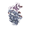
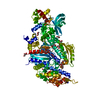
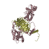
Entry Database : PDB / ID : 2pbiTitle The multifunctional nature of Gbeta5/RGS9 revealed from its crystal structure Guanine nucleotide-binding protein subunit beta 5 Regulator of G-protein signaling 9 Keywords / / / / / / Function / homology Function Domain/homology Component
/ / / / / / / / / / / / / / / / / / / / / / / / / / / / / / / / / / / / / / / / / / / / / / / / / / / / / / / / / / / / / / / / / / / / / / / / / / / / / / / / / / / / / / / / / / / / / / / / / / / / / / / / / / / / / / / / / / / / / / / / / / / / / / / / / / / / / / Biological species Mus musculus (house mouse)Method / / / Resolution : 1.95 Å Authors Cheever, M.L. / Snyder, J.T. / Gershburg, S. / Siderovski, D.P. / Harden, T.K. / Sondek, J. Journal : Nat.Struct.Mol.Biol. / Year : 2008Title : Crystal structure of the multifunctional Gbeta5-RGS9 complex.Authors : Cheever, M.L. / Snyder, J.T. / Gershburg, S. / Siderovski, D.P. / Harden, T.K. / Sondek, J. History Deposition Mar 28, 2007 Deposition site / Processing site Revision 1.0 Jan 29, 2008 Provider / Type Revision 1.1 Jul 13, 2011 Group / Version format complianceRevision 1.2 Oct 18, 2017 Group / Category Item _software.classification / _software.contact_author ... _software.classification / _software.contact_author / _software.contact_author_email / _software.date / _software.language / _software.location / _software.name / _software.type / _software.version Revision 1.3 Feb 21, 2024 Group / Database references / Derived calculationsCategory chem_comp_atom / chem_comp_bond ... chem_comp_atom / chem_comp_bond / database_2 / struct_ref_seq_dif / struct_site Item _database_2.pdbx_DOI / _database_2.pdbx_database_accession ... _database_2.pdbx_DOI / _database_2.pdbx_database_accession / _struct_ref_seq_dif.details / _struct_site.pdbx_auth_asym_id / _struct_site.pdbx_auth_comp_id / _struct_site.pdbx_auth_seq_id
Show all Show less
 Movie
Movie Controller

 Yorodumi
 Open data
 Basic information
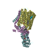
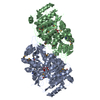
 Components
 Keywords
 Function and homology information
 X-RAY DIFFRACTION /
  Authors
 Citation
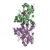
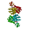
 Structure visualization
 Downloads & links
 Other downloads
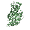

 PDBj
PDBj


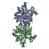
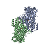
 Assembly

 Trichoplusia ni (cabbage looper) / Strain (production host): Hi5 / References: UniProt: A1L352, UniProt: O54828*PLUS
 Mass: 92.094 Da / Num. of mol.: 9 / Source method: obtained synthetically / Formula: C3H8O3
 Mass: 18.015 Da / Num. of mol.: 890 / Source method: isolated from a natural source / Formula: H2O
 Sample preparation
 / Beamline: 22-ID / Wavelength: 1 Å
 Processing