 Movie
Movie Controller

[English] 日本語
 Yorodumi
- PDB-6qdj: Molecular features of the UNC-45 chaperone critical for binding a... -
+ Open data

- Basic information
| Entry | Database: PDB / ID: 6qdj | ||||||
|---|---|---|---|---|---|---|---|
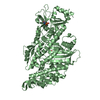
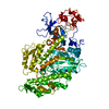
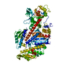
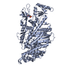
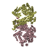
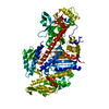
| Title | Molecular features of the UNC-45 chaperone critical for binding and folding muscle myosin | ||||||
 Components Components | Myosin-4 | ||||||
 Keywords Keywords | MOTOR PROTEIN / MYOSIN / MHC-B / UNC-54 | ||||||
| Function / homology |  Function and homology information Function and homology informationegg-laying behavior / striated muscle myosin thick filament / skeletal muscle myosin thick filament assembly / A band / muscle myosin complex / myosin filament / locomotion / myosin II complex / sarcomere organization / structural constituent of muscle ...egg-laying behavior / striated muscle myosin thick filament / skeletal muscle myosin thick filament assembly / A band / muscle myosin complex / myosin filament / locomotion / myosin II complex / sarcomere organization / structural constituent of muscle / microfilament motor activity / muscle contraction / actin filament binding / ATP binding / cytoplasm Similarity search - Function | ||||||
| Biological species |  | ||||||
| Method |  X-RAY DIFFRACTION / SYNCHROTRON / MOLECULAR REPLACEMENT / Resolution: 1.884 Å X-RAY DIFFRACTION / SYNCHROTRON / MOLECULAR REPLACEMENT / Resolution: 1.884 Å | ||||||
 Authors Authors | Meinhart, A. / Clausen, T. / Arnese, R. | ||||||
| Funding support |  Austria, 1items Austria, 1items
| ||||||
 Citation Citation | Journal: Nat Commun / Year: 2019 Title: Molecular features of the UNC-45 chaperone critical for binding and folding muscle myosin. Authors: Hellerschmied, D. / Lehner, A. / Franicevic, N. / Arnese, R. / Johnson, C. / Vogel, A. / Meinhart, A. / Kurzbauer, R. / Deszcz, L. / Gazda, L. / Geeves, M. / Clausen, T. | ||||||
| History |
|
- Structure visualization
| Structure viewer | Molecule: MolmilJmol/JSmol |
|---|
- Downloads & links
-Download
| PDBx/mmCIF format | 6qdj.cif.gz | 178.5 KB | Display | PDBx/mmCIF format |
|---|---|---|---|---|
| PDB format | pdb6qdj.ent.gz | 136.9 KB | Display | PDB format |
| PDBx/mmJSON format | 6qdj.json.gz | Tree view | PDBx/mmJSON format | |
| Others |  Other downloads Other downloads |
-Validation report
| Arichive directory | https://data.pdbj.org/pub/pdb/validation_reports/qd/6qdjftp://data.pdbj.org/pub/pdb/validation_reports/qd/6qdj | HTTPS FTP |
|---|
-Related structure data
| Related structure data | 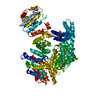 6qdkC 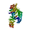 6qdlC  6qdmC 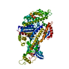 5m05S S: Starting model for refinement C: citing same article ( |
|---|---|
| Similar structure data |


-Links
 PDBj
PDBj


- Assembly
| Deposited unit | 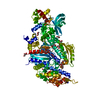
| ||||||||
|---|---|---|---|---|---|---|---|---|---|
| 1 |
| ||||||||
| Unit cell |
|
-Components
-Protein , 1 types, 1 molecules A
| #1: Protein | Mass: 90372.297 Da / Num. of mol.: 1 Source method: isolated from a genetically manipulated source Source: (gene. exp.)  Trichoplusia ni (cabbage looper) / References: UniProt: P02566 Trichoplusia ni (cabbage looper) / References: UniProt: P02566 |
|---|
-Non-polymers , 7 types, 253 molecules 


| #2: Chemical | ChemComp-ADP /  Mass: 427.201 Da / Num. of mol.: 1 / Source method: obtained synthetically / Formula: C10H15N5O10P2 / Comment: ADP, energy-carrying molecule*YM Mass: 427.201 Da / Num. of mol.: 1 / Source method: obtained synthetically / Formula: C10H15N5O10P2 / Comment: ADP, energy-carrying molecule*YM | ||||||||||
|---|---|---|---|---|---|---|---|---|---|---|---|
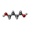
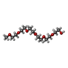
| #3: Chemical | ChemComp-GOL /  Mass: 92.094 Da / Num. of mol.: 10 / Source method: obtained synthetically / Formula: C3H8O3 Mass: 92.094 Da / Num. of mol.: 10 / Source method: obtained synthetically / Formula: C3H8O3#4: Chemical | ChemComp-BU1 / |  Mass: 90.121 Da / Num. of mol.: 1 / Source method: obtained synthetically / Formula: C4H10O2 Mass: 90.121 Da / Num. of mol.: 1 / Source method: obtained synthetically / Formula: C4H10O2#5: Chemical |  Mass: 76.094 Da / Num. of mol.: 2 / Source method: obtained synthetically / Formula: C3H8O2 Mass: 76.094 Da / Num. of mol.: 2 / Source method: obtained synthetically / Formula: C3H8O2#6: Chemical | ChemComp-HEZ / |  Mass: 118.174 Da / Num. of mol.: 1 / Source method: obtained synthetically / Formula: C6H14O2 Mass: 118.174 Da / Num. of mol.: 1 / Source method: obtained synthetically / Formula: C6H14O2#7: Chemical | ChemComp-P15 / |  Mass: 296.357 Da / Num. of mol.: 1 / Source method: obtained synthetically / Formula: C13H28O7 Mass: 296.357 Da / Num. of mol.: 1 / Source method: obtained synthetically / Formula: C13H28O7#8: Water | ChemComp-HOH / | Mass: 18.015 Da / Num. of mol.: 237 / Source method: isolated from a natural source / Formula: H2O |
-Experimental details
-Experiment
| Experiment | Method: X-RAY DIFFRACTION / Number of used crystals: 1 |
|---|
- Sample preparation
| Crystal | Density Matthews: 2.82 Å3/Da / Density % sol: 56.33 % |
|---|---|
| Crystal grow | Temperature: 277 K / Method: vapor diffusion, sitting drop / pH: 6.5 Details: 100 mM MES/imidazole pH 6.5 10 % PEG 4000 20 % glycerol 20 mM 1,6-hexandiol 20 mM 1-butanol 20 mM (RS)-1,2-propanediol 20 mM 2-propanol 20 mM 1,4-butanediol 20 mM 1,3-propandiol |
-Data collection
| Diffraction | Mean temperature: 100 K / Serial crystal experiment: N |
|---|---|
| Diffraction source | Source: SYNCHROTRON / Site: PETRA III, EMBL c/o DESY  / Beamline: P13 (MX1) / Wavelength: 0.9763 Å / Beamline: P13 (MX1) / Wavelength: 0.9763 Å |
| Detector | Type: DECTRIS PILATUS 6M / Detector: PIXEL / Date: Dec 17, 2016 |
| Radiation | Monochromator: KB-mirror / Protocol: SINGLE WAVELENGTH / Monochromatic (M) / Laue (L): M / Scattering type: x-ray |
| Radiation wavelength | Wavelength: 0.9763 Å / Relative weight: 1 |
| Reflection | Resolution: 1.884→85 Å / Num. obs: 76208 / % possible obs: 93.8 % / Redundancy: 3.3 % / Biso Wilson estimate: 37.5 Å2 / CC1/2: 0.999 / Rrim(I) all: 0.045 / Net I/σ(I): 19.6 |
| Reflection shell | Resolution: 1.884→2.05 Å / Redundancy: 2.9 % / Mean I/σ(I) obs: 2.9 / CC1/2: 0.866 / Rrim(I) all: 0.454 / % possible all: 78 |
- Processing
| Software |
| ||||||||||||||||||||||||||||||||||||||||||||||||||||||||||||||||||||||||||||||||||||||||||||||||||||||||||||||||||||||||||||||||||||||||||||||||||||||||||||||||||||||||||||||||||||||||||||||||||||
|---|---|---|---|---|---|---|---|---|---|---|---|---|---|---|---|---|---|---|---|---|---|---|---|---|---|---|---|---|---|---|---|---|---|---|---|---|---|---|---|---|---|---|---|---|---|---|---|---|---|---|---|---|---|---|---|---|---|---|---|---|---|---|---|---|---|---|---|---|---|---|---|---|---|---|---|---|---|---|---|---|---|---|---|---|---|---|---|---|---|---|---|---|---|---|---|---|---|---|---|---|---|---|---|---|---|---|---|---|---|---|---|---|---|---|---|---|---|---|---|---|---|---|---|---|---|---|---|---|---|---|---|---|---|---|---|---|---|---|---|---|---|---|---|---|---|---|---|---|---|---|---|---|---|---|---|---|---|---|---|---|---|---|---|---|---|---|---|---|---|---|---|---|---|---|---|---|---|---|---|---|---|---|---|---|---|---|---|---|---|---|---|---|---|---|---|---|---|
| Refinement | Method to determine structure: MOLECULAR REPLACEMENT Starting model: 5m05 Resolution: 1.884→84.026 Å / SU ML: 0.19 / Cross valid method: FREE R-VALUE / σ(F): 1.37 / Phase error: 22.81
| ||||||||||||||||||||||||||||||||||||||||||||||||||||||||||||||||||||||||||||||||||||||||||||||||||||||||||||||||||||||||||||||||||||||||||||||||||||||||||||||||||||||||||||||||||||||||||||||||||||
| Solvent computation | Shrinkage radii: 0.9 Å / VDW probe radii: 1.11 Å | ||||||||||||||||||||||||||||||||||||||||||||||||||||||||||||||||||||||||||||||||||||||||||||||||||||||||||||||||||||||||||||||||||||||||||||||||||||||||||||||||||||||||||||||||||||||||||||||||||||
| Refinement step | Cycle: LAST / Resolution: 1.884→84.026 Å
| ||||||||||||||||||||||||||||||||||||||||||||||||||||||||||||||||||||||||||||||||||||||||||||||||||||||||||||||||||||||||||||||||||||||||||||||||||||||||||||||||||||||||||||||||||||||||||||||||||||
| Refine LS restraints |
| ||||||||||||||||||||||||||||||||||||||||||||||||||||||||||||||||||||||||||||||||||||||||||||||||||||||||||||||||||||||||||||||||||||||||||||||||||||||||||||||||||||||||||||||||||||||||||||||||||||
| LS refinement shell |
|
