 Movie
Movie Controller

[English] 日本語
 Yorodumi
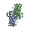
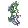
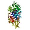
- PDB-6vzy: Crystal structure of SznF from Streptomyces achromogenes var. str... -
+ Open data

- Basic information
| Entry | Database: PDB / ID: 6vzy | ||||||
|---|---|---|---|---|---|---|---|
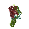
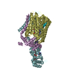
| Title | Crystal structure of SznF from Streptomyces achromogenes var. streptozoticus NRRL 2697 with a diiron(II) central domain cofactor | ||||||
 Components Components | Cupin domain-containing diiron protein | ||||||
 Keywords Keywords | OXIDOREDUCTASE / diiron / N-oxygenase / N-methyl-L-arginine / heme oxygenase | ||||||
| Function / homology |  Function and homology information Function and homology informationnitrosourea synthase / antibiotic biosynthetic process / monooxygenase activity / metal ion binding Similarity search - Function | ||||||
| Biological species |  Streptomyces achromogenes subsp. streptozoticus (bacteria) Streptomyces achromogenes subsp. streptozoticus (bacteria) | ||||||
| Method |  X-RAY DIFFRACTION / SYNCHROTRON / MOLECULAR REPLACEMENT / Resolution: 1.66 Å X-RAY DIFFRACTION / SYNCHROTRON / MOLECULAR REPLACEMENT / Resolution: 1.66 Å | ||||||
 Authors Authors | McBride, M.J. / Boal, A.K. | ||||||
| Funding support | 1items
| ||||||
 Citation Citation | Journal: Proc.Natl.Acad.Sci.USA / Year: 2021 Title: Structure and assembly of the diiron cofactor in the heme-oxygenase-like domain of the N -nitrosourea-producing enzyme SznF. Authors: McBride, M.J. / Pope, S.R. / Hu, K. / Okafor, C.D. / Balskus, E.P. / Bollinger Jr., J.M. / Boal, A.K. | ||||||
| History |
|
- Structure visualization
| Structure viewer | Molecule: MolmilJmol/JSmol |
|---|
- Downloads & links
-Download
| PDBx/mmCIF format | 6vzy.cif.gz | 240.9 KB | Display | PDBx/mmCIF format |
|---|---|---|---|---|
| PDB format | pdb6vzy.ent.gz | 161.7 KB | Display | PDB format |
| PDBx/mmJSON format | 6vzy.json.gz | Tree view | PDBx/mmJSON format | |
| Others |  Other downloads Other downloads |
-Validation report
| Arichive directory | https://data.pdbj.org/pub/pdb/validation_reports/vz/6vzyftp://data.pdbj.org/pub/pdb/validation_reports/vz/6vzy | HTTPS FTP |
|---|
-Related structure data
| Related structure data | 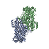 6xcvC  6m9rS S: Starting model for refinement C: citing same article ( |
|---|---|
| Similar structure data |


-Links
 PDBj
PDBj


- Assembly
| Deposited unit | 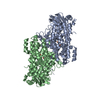
| ||||||||||||
|---|---|---|---|---|---|---|---|---|---|---|---|---|---|
| 1 |
| ||||||||||||
| Unit cell |
|
-Components
| #1: Protein | Mass: 56042.543 Da / Num. of mol.: 2 Source method: isolated from a genetically manipulated source Source: (gene. exp.) Streptomyces achromogenes subsp. streptozoticus (bacteria)Strain: NRRL 2697 / Gene: stzF, sznF / Production host: #2: Chemical | ChemComp-FE2 /   Mass: 55.845 Da / Num. of mol.: 6 / Source method: obtained synthetically / Formula: Fe / Feature type: SUBJECT OF INVESTIGATION Mass: 55.845 Da / Num. of mol.: 6 / Source method: obtained synthetically / Formula: Fe / Feature type: SUBJECT OF INVESTIGATION#3: Water | ChemComp-HOH / |  Mass: 18.015 Da / Num. of mol.: 411 / Source method: isolated from a natural source / Formula: H2O Mass: 18.015 Da / Num. of mol.: 411 / Source method: isolated from a natural source / Formula: H2OHas ligand of interest | Y | |
|---|
-Experimental details
-Experiment
| Experiment | Method: X-RAY DIFFRACTION / Number of used crystals: 1 |
|---|
- Sample preparation
| Crystal | Density Matthews: 2.06 Å3/Da / Density % sol: 40.3 % |
|---|---|
| Crystal grow | Temperature: 298 K / Method: vapor diffusion, hanging drop Details: 16% PEG4000, 0.1 M magnesium chloride, 0.1 M Tris, pH 8.5 |
-Data collection
| Diffraction | Mean temperature: 100 K / Serial crystal experiment: N |
|---|---|
| Diffraction source | Source: SYNCHROTRON / Site: APS  / Beamline: 23-ID-B / Wavelength: 1.03319 Å / Beamline: 23-ID-B / Wavelength: 1.03319 Å |
| Detector | Type: DECTRIS EIGER X 16M / Detector: PIXEL / Date: Nov 13, 2019 |
| Radiation | Monochromator: Double crystal cryo-cooled Si(111) / Protocol: SINGLE WAVELENGTH / Monochromatic (M) / Laue (L): M / Scattering type: x-ray |
| Radiation wavelength | Wavelength: 1.03319 Å / Relative weight: 1 |
| Reflection | Resolution: 1.66→50 Å / Num. obs: 109965 / % possible obs: 96.8 % / Redundancy: 5.8 % / Biso Wilson estimate: 14.43 Å2 / CC1/2: 0.996 / Net I/σ(I): 23.6 |
| Reflection shell | Resolution: 1.66→1.69 Å / Num. unique obs: 109902 / CC1/2: 0.677 |
- Processing
| Software |
| ||||||||||||||||||
|---|---|---|---|---|---|---|---|---|---|---|---|---|---|---|---|---|---|---|---|
| Refinement | Method to determine structure: MOLECULAR REPLACEMENT Starting model: PDB entry 6M9R Resolution: 1.66→36.42 Å / Cross valid method: FREE R-VALUE / σ(F): 1.34
| ||||||||||||||||||
| Displacement parameters | Biso mean: 18.14 Å2 | ||||||||||||||||||
| Refinement step | Cycle: LAST / Resolution: 1.66→36.42 Å
| ||||||||||||||||||
| LS refinement shell | Resolution: 1.66→1.72 Å
|
