 Movie
Movie Controller

[English] 日本語
 Yorodumi
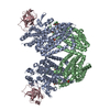
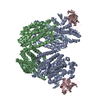
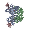
- PDB-3n1x: X-ray Crystal Structure of Toluene/o-Xylene Monooxygenase Hydroxy... -
+ Open data

- Basic information
| Entry | Database: PDB / ID: 3n1x | ||||||
|---|---|---|---|---|---|---|---|
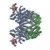
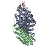
| Title | X-ray Crystal Structure of Toluene/o-Xylene Monooxygenase Hydroxylase T201C Mutant | ||||||
 Components Components | (Toluene o-xylene monooxygenase ...) x 3 | ||||||
 Keywords Keywords | OXIDOREDUCTASE / Diiron / 4-helix bundle / carboxylate bridge / metalloenzyme | ||||||
| Function / homology |  Function and homology information Function and homology informationoxidoreductase activity, acting on paired donors, with incorporation or reduction of molecular oxygen, NAD(P)H as one donor, and incorporation of one atom of oxygen / monooxygenase activity / metal ion binding Similarity search - Function | ||||||
| Biological species |  Pseudomonas sp. (bacteria) Pseudomonas sp. (bacteria) | ||||||
| Method |  X-RAY DIFFRACTION / SYNCHROTRON / MOLECULAR REPLACEMENT / Resolution: 2.4 Å X-RAY DIFFRACTION / SYNCHROTRON / MOLECULAR REPLACEMENT / Resolution: 2.4 Å | ||||||
 Authors Authors | Sazinsky, M.H. / McCormick, M.S. / Lippard, S.J. | ||||||
 Citation Citation | Journal: J.Am.Chem.Soc. / Year: 2010 Title: Active Site Threonine Facilitates Proton Transfer during Dioxygen Activation at the Diiron Center of Toluene/o-Xylene Monooxygenase Hydroxylase. Authors: Song, W.J. / McCormick, M.S. / Behan, R.K. / Sazinsky, M.H. / Jiang, W. / Lin, J. / Krebs, C. / Lippard, S.J. | ||||||
| History |
|
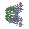
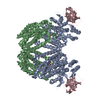
- Structure visualization
| Structure viewer | Molecule: MolmilJmol/JSmol |
|---|
- Downloads & links
-Download
| PDBx/mmCIF format | 3n1x.cif.gz | 199.1 KB | Display | PDBx/mmCIF format |
|---|---|---|---|---|
| PDB format | pdb3n1x.ent.gz | 156.3 KB | Display | PDB format |
| PDBx/mmJSON format | 3n1x.json.gz | Tree view | PDBx/mmJSON format | |
| Others |  Other downloads Other downloads |
-Validation report
| Arichive directory | https://data.pdbj.org/pub/pdb/validation_reports/n1/3n1xftp://data.pdbj.org/pub/pdb/validation_reports/n1/3n1x | HTTPS FTP |
|---|
-Related structure data
| Related structure data |  3n1yC 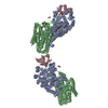 3n1zC  3n20C 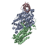 2incS S: Starting model for refinement C: citing same article ( |
|---|---|
| Similar structure data |


-Links
 PDBj
PDBj


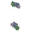
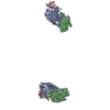
- Assembly
| Deposited unit | 
| ||||||||
|---|---|---|---|---|---|---|---|---|---|
| 1 | 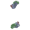
| ||||||||
| 2 |
| ||||||||
| Unit cell |
|
-Components
-Toluene o-xylene monooxygenase ... , 3 types, 3 molecules ABC
| #1: Protein | Mass: 57788.680 Da / Num. of mol.: 1 / Mutation: T201C Source method: isolated from a genetically manipulated source Source: (gene. exp.) Pseudomonas sp. (bacteria) / Strain: OX1 / Gene: touA / Production host: |
|---|---|
| #2: Protein | Mass: 38381.059 Da / Num. of mol.: 1 Source method: isolated from a genetically manipulated source Source: (gene. exp.) Pseudomonas sp. (bacteria) / Strain: OX1 / Gene: touE / Production host: |
| #3: Protein | Mass: 9986.430 Da / Num. of mol.: 1 Source method: isolated from a genetically manipulated source Source: (gene. exp.) Pseudomonas sp. (bacteria) / Strain: OX1 / Gene: touB / Production host: |
-Non-polymers , 5 types, 145 molecules 


| #4: Chemical |  Mass: 55.845 Da / Num. of mol.: 2 / Source method: obtained synthetically / Formula: Fe Mass: 55.845 Da / Num. of mol.: 2 / Source method: obtained synthetically / Formula: Fe#5: Chemical | ChemComp-OH / |  Mass: 17.007 Da / Num. of mol.: 1 / Source method: obtained synthetically / Formula: HO Mass: 17.007 Da / Num. of mol.: 1 / Source method: obtained synthetically / Formula: HO#6: Chemical | ChemComp-EDO / |  Mass: 62.068 Da / Num. of mol.: 1 / Source method: obtained synthetically / Formula: C2H6O2 Mass: 62.068 Da / Num. of mol.: 1 / Source method: obtained synthetically / Formula: C2H6O2#7: Chemical | ChemComp-SO4 / |  Mass: 96.063 Da / Num. of mol.: 1 / Source method: obtained synthetically / Formula: SO4 Mass: 96.063 Da / Num. of mol.: 1 / Source method: obtained synthetically / Formula: SO4#8: Water | ChemComp-HOH / | Mass: 18.015 Da / Num. of mol.: 140 / Source method: isolated from a natural source / Formula: H2O |
|---|
-Experimental details
-Experiment
| Experiment | Method: X-RAY DIFFRACTION / Number of used crystals: 1 |
|---|
- Sample preparation
| Crystal | Density Matthews: 3.11 Å3/Da / Density % sol: 60.48 % |
|---|---|
| Crystal grow | Temperature: 293 K / Method: vapor diffusion, hanging drop / pH: 7.5 Details: 100 mM HEPES pH 7.5, 2.1-2.5 M Ammonium Sulfate, 2-4% PEG 400, VAPOR DIFFUSION, HANGING DROP, temperature 293K |
-Data collection
| Diffraction | Mean temperature: 100 K |
|---|---|
| Diffraction source | Source: SYNCHROTRON / Site: SSRL  / Beamline: BL11-1 / Wavelength: 0.979 Å / Beamline: BL11-1 / Wavelength: 0.979 Å |
| Detector | Type: MARMOSAIC 325 mm CCD / Detector: CCD / Date: Feb 11, 2008 / Details: Rh coated flat mirror |
| Radiation | Monochromator: Side scattering bent cube-root I-beam single crystal; asymmetric cut 4.965 degrees Protocol: SINGLE WAVELENGTH / Monochromatic (M) / Laue (L): M / Scattering type: x-ray |
| Radiation wavelength | Wavelength: 0.979 Å / Relative weight: 1 |
| Reflection | Resolution: 2.4→50 Å / Num. all: 48929 / Num. obs: 48523 / % possible obs: 99.17 % / Observed criterion σ(F): 0 / Observed criterion σ(I): 0 |
| Reflection shell | Resolution: 2.4→2.462 Å / % possible all: 96.16 |
- Processing
| Software |
| |||||||||||||||||||||||||
|---|---|---|---|---|---|---|---|---|---|---|---|---|---|---|---|---|---|---|---|---|---|---|---|---|---|---|
| Refinement | Method to determine structure: MOLECULAR REPLACEMENT Starting model: PDB ENTRY 2INC Resolution: 2.4→38.02 Å / σ(F): 0 / σ(I): 0 / Stereochemistry target values: Engh & Huber
| |||||||||||||||||||||||||
| Refinement step | Cycle: LAST / Resolution: 2.4→38.02 Å
| |||||||||||||||||||||||||
| Refine LS restraints |
|
