 Movie
Movie Controller

[English] 日本語
 Yorodumi
- PDB-1xr4: X-ray crystal structure of putative citrate lyase alpha chain/cit... -
+ Open data

- Basic information
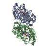
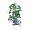
| Entry | Database: PDB / ID: 1xr4 | ||||||
|---|---|---|---|---|---|---|---|
| Title | X-ray crystal structure of putative citrate lyase alpha chain/citrate-ACP transferase [Salmonella typhimurium] | ||||||
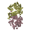
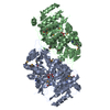
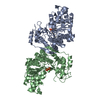
 Components Components | putative citrate lyase alpha chain/citrate-ACP transferase | ||||||
 Keywords Keywords | HYDROLASE/TRANSFERASE / The Midwest Center for Structural Genomics / MCSG / structural genomics / protein structure initiative / PSI / citrate lyase / HYDROLASE-TRANSFERASE COMPLEX | ||||||
| Function / homology |  Function and homology information Function and homology informationcitrate CoA-transferase / citrate CoA-transferase activity / ATP-independent citrate lyase complex / citrate (pro-3S)-lyase / citrate (pro-3S)-lyase activity / citrate metabolic process / acetyl-CoA metabolic process / cytoplasm Similarity search - Function | ||||||
| Biological species |  Salmonella typhimurium (bacteria) Salmonella typhimurium (bacteria) | ||||||
| Method |  X-RAY DIFFRACTION / SYNCHROTRON / SAD / Resolution: 2.37 Å X-RAY DIFFRACTION / SYNCHROTRON / SAD / Resolution: 2.37 Å | ||||||
 Authors Authors | Osipiuk, J. / Quartey, P. / Moy, S. / Collart, F. / Joachimiak, A. / Midwest Center for Structural Genomics (MCSG) | ||||||
 Citation Citation | Journal: To be Published Title: X-ray crystal structure of putative citrate lyase alpha chain/citrate-ACP transferase from Salmonella typhimurium. Authors: Osipiuk, J. / Quartey, P. / Moy, S. / Collart, F. / Joachimiak, A. | ||||||
| History |
| ||||||
| Remark 300 | BIOMOLECULE: 1 THIS ENTRY CONTAINS THE CRYSTALLOGRAPHIC ASYMMETRIC UNIT WHICH CONSISTS OF 2 CHAIN(S) ...BIOMOLECULE: 1 THIS ENTRY CONTAINS THE CRYSTALLOGRAPHIC ASYMMETRIC UNIT WHICH CONSISTS OF 2 CHAIN(S). THE BIOLOGICAL MOLECULE FOR THE PROTEIN IS UNKNOWN. |
- Structure visualization
| Structure viewer | Molecule: MolmilJmol/JSmol |
|---|
- Downloads & links
-Download
| PDBx/mmCIF format | 1xr4.cif.gz | 203.1 KB | Display | PDBx/mmCIF format |
|---|---|---|---|---|
| PDB format | pdb1xr4.ent.gz | 163.6 KB | Display | PDB format |
| PDBx/mmJSON format | 1xr4.json.gz | Tree view | PDBx/mmJSON format | |
| Others |  Other downloads Other downloads |
-Validation report
| Arichive directory | https://data.pdbj.org/pub/pdb/validation_reports/xr/1xr4ftp://data.pdbj.org/pub/pdb/validation_reports/xr/1xr4 | HTTPS FTP |
|---|
-Related structure data
| Similar structure data | |
|---|---|
| Other databases |


-Links
 PDBj
PDBj- Assembly
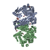
| Deposited unit | 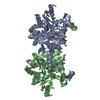
| ||||||||
|---|---|---|---|---|---|---|---|---|---|
| 1 |
| ||||||||
| Unit cell |
|
-Components
| #1: Protein | Mass: 55445.934 Da / Num. of mol.: 2 Source method: isolated from a genetically manipulated source Source: (gene. exp.) Salmonella typhimurium (bacteria) / Strain: LT2 / Gene: CitF / Plasmid: pMCSG7 / Species (production host): Escherichia coli / Production host: #2: Water | ChemComp-HOH / |  Mass: 18.015 Da / Num. of mol.: 287 / Source method: isolated from a natural source / Formula: H2O Mass: 18.015 Da / Num. of mol.: 287 / Source method: isolated from a natural source / Formula: H2OHas protein modification | Y | |
|---|
-Experimental details
-Experiment
| Experiment | Method: X-RAY DIFFRACTION / Number of used crystals: 1 |
|---|
- Sample preparation
| Crystal | Density Matthews: 2.9 Å3/Da / Density % sol: 57.9 % |
|---|---|
| Crystal grow | Temperature: 294 K / Method: vapor diffusion, hanging drop / pH: 8.5 Details: Tris_HCl, PEG 8000, pH 8.5, VAPOR DIFFUSION, HANGING DROP, temperature 294K |
-Data collection
| Diffraction | Mean temperature: 100 K |
|---|---|
| Diffraction source | Source: SYNCHROTRON / Site: APS  / Beamline: 19-BM / Wavelength: 0.979565 Å / Beamline: 19-BM / Wavelength: 0.979565 Å |
| Detector | Type: SBC-3 / Detector: CCD / Date: Mar 27, 2004 |
| Radiation | Monochromator: double crystal monochromator / Protocol: SINGLE WAVELENGTH / Monochromatic (M) / Laue (L): M / Scattering type: x-ray |
| Radiation wavelength | Wavelength: 0.979565 Å / Relative weight: 1 |
| Reflection | Resolution: 2.37→40 Å / Num. all: 53458 / Num. obs: 49002 / % possible obs: 91.7 % / Observed criterion σ(F): 0 / Observed criterion σ(I): 0 / Redundancy: 12.1 % / Rmerge(I) obs: 0.184 / Net I/σ(I): 13.34 |
| Reflection shell | Resolution: 2.4→2.47 Å / Redundancy: 5 % / Rmerge(I) obs: 0.524 / Mean I/σ(I) obs: 1.85 / Num. unique all: 2256 / % possible all: 51.5 |
- Processing
| Software |
| ||||||||||||||||||||||||||||||||||||||||||||||||||||||||||||||||||||||||||||||||||||||||||||||||||||||||||||||||||||||||||||||||||||||||||||||||||||||||||||||||
|---|---|---|---|---|---|---|---|---|---|---|---|---|---|---|---|---|---|---|---|---|---|---|---|---|---|---|---|---|---|---|---|---|---|---|---|---|---|---|---|---|---|---|---|---|---|---|---|---|---|---|---|---|---|---|---|---|---|---|---|---|---|---|---|---|---|---|---|---|---|---|---|---|---|---|---|---|---|---|---|---|---|---|---|---|---|---|---|---|---|---|---|---|---|---|---|---|---|---|---|---|---|---|---|---|---|---|---|---|---|---|---|---|---|---|---|---|---|---|---|---|---|---|---|---|---|---|---|---|---|---|---|---|---|---|---|---|---|---|---|---|---|---|---|---|---|---|---|---|---|---|---|---|---|---|---|---|---|---|---|---|---|
| Refinement | Method to determine structure: SAD / Resolution: 2.37→40 Å / Cor.coef. Fo:Fc: 0.946 / SU B: 11.417 / SU ML: 0.134 / TLS residual ADP flag: LIKELY RESIDUAL / σ(F): 0 / σ(I): 0 / ESU R: 0.316 / Stereochemistry target values: MAXIMUM LIKELIHOOD Details: HYDROGENS HAVE BEEN ADDED IN THE RIDING POSITIONS. The test data set was not used in last round of refinement. The deposited pdb file is from the last round of refinement. R-factor-obs and R- ...Details: HYDROGENS HAVE BEEN ADDED IN THE RIDING POSITIONS. The test data set was not used in last round of refinement. The deposited pdb file is from the last round of refinement. R-factor-obs and R-factor-all correspond to deposited file. R-factor-work and R-factor-free are taken from second to last round of refinement which used test data set.
| ||||||||||||||||||||||||||||||||||||||||||||||||||||||||||||||||||||||||||||||||||||||||||||||||||||||||||||||||||||||||||||||||||||||||||||||||||||||||||||||||
| Solvent computation | Ion probe radii: 0.8 Å / Shrinkage radii: 0.8 Å / VDW probe radii: 1.2 Å / Solvent model: MASK | ||||||||||||||||||||||||||||||||||||||||||||||||||||||||||||||||||||||||||||||||||||||||||||||||||||||||||||||||||||||||||||||||||||||||||||||||||||||||||||||||
| Displacement parameters | Biso mean: 35.982 Å2
| ||||||||||||||||||||||||||||||||||||||||||||||||||||||||||||||||||||||||||||||||||||||||||||||||||||||||||||||||||||||||||||||||||||||||||||||||||||||||||||||||
| Refinement step | Cycle: LAST / Resolution: 2.37→40 Å
| ||||||||||||||||||||||||||||||||||||||||||||||||||||||||||||||||||||||||||||||||||||||||||||||||||||||||||||||||||||||||||||||||||||||||||||||||||||||||||||||||
| Refine LS restraints |
| ||||||||||||||||||||||||||||||||||||||||||||||||||||||||||||||||||||||||||||||||||||||||||||||||||||||||||||||||||||||||||||||||||||||||||||||||||||||||||||||||
| LS refinement shell | Resolution: 2.373→2.435 Å / Total num. of bins used: 20
| ||||||||||||||||||||||||||||||||||||||||||||||||||||||||||||||||||||||||||||||||||||||||||||||||||||||||||||||||||||||||||||||||||||||||||||||||||||||||||||||||
| Refinement TLS params. | Method: refined / Refine-ID: X-RAY DIFFRACTION
| ||||||||||||||||||||||||||||||||||||||||||||||||||||||||||||||||||||||||||||||||||||||||||||||||||||||||||||||||||||||||||||||||||||||||||||||||||||||||||||||||
| Refinement TLS group |
|
