 Movie
Movie Controller

[English] 日本語
 Yorodumi
- PDB-1rb8: The phiX174 DNA binding protein J in two different capsid environ... -
+ Open data

- Basic information
| Entry | Database: PDB / ID: 1rb8 | |||||||||
|---|---|---|---|---|---|---|---|---|---|---|
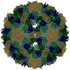
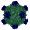
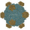
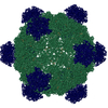
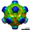
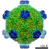
| Title | The phiX174 DNA binding protein J in two different capsid environments. | |||||||||
 Components Components |
| |||||||||
 Keywords Keywords | Virus/DNA / bacteriophage alpha3 / bacteriophage phiX174 / bacteriophage alpha3 chimera / alpha3 / phiX174 / three-dimentional structure / virion / Microviridae / Icosahedral virus / Virus-DNA COMPLEX | |||||||||
| Function / homology |  Function and homology information Function and homology informationsymbiont-mediated perturbation of host process / T=1 icosahedral viral capsid / viral capsid / host cell cytoplasm / symbiont entry into host cell / virion attachment to host cell / structural molecule activity / DNA binding Similarity search - Function | |||||||||
| Biological species |  Enterobacteria phage phiX174 (virus)Enterobacteria phage alpha3 (virus) Enterobacteria phage phiX174 (virus)Enterobacteria phage alpha3 (virus) | |||||||||
| Method |  X-RAY DIFFRACTION / SYNCHROTRON / MOLECULAR REPLACEMENT / Resolution: 3.5 Å X-RAY DIFFRACTION / SYNCHROTRON / MOLECULAR REPLACEMENT / Resolution: 3.5 Å | |||||||||
 Authors Authors | Bernal, R.A. / Hafenstein, S. / Esmeralda, R. / Fane, B.A. / Rossmann, M.G. | |||||||||
 Citation Citation | Journal: J.Mol.Biol. / Year: 2004 Title: The phiX174 Protein J Mediates DNA Packaging and Viral Attachment to Host Cells. Authors: Bernal, R.A. / Hafenstein, S. / Esmeralda, R. / Fane, B.A. / Rossmann, M.G. #1: Journal: J.Mol.Biol. / Year: 2003Title: Structural Studies of Bacteriophage Alpha3 Assembly Authors: Bernal, R.A. / Hafenstein, S. / Olson, N.H. / Bowman, V.D. / Chipman, P.R. / Baker, T.S. / Fane, B.A. / Rossmann, M.G. #2: Journal: J.Mol.Biol. / Year: 1999Title: The role of scaffolding proteins in the assembly of the small, single-stranded DNA virus phiX174. Authors: Dokland, T. / Bernal, R.A. / Burch, A. / Pletnev, S. / Fane, B.A. / Rossmann, M.G. #3: Journal: Nature / Year: 1992Title: Atomic structure of single-stranded DNA bacteriophage phi X174 and its functional implications. Authors: McKenna, R. / Xia, D. / Willingmann, P. / Ilag, L.L. / Krishnaswamy, S. / Rossmann, M.G. / Olson, N.H. / Baker, T.S. / Incardona, N.L. | |||||||||
| History |
| |||||||||
| Remark 400 | COMPOUND THE VIRUS IS A CHIMERA. THE CAPSID PROTEIN F AND THE SPIKE PROTEIN G ARE FROM ALPHA3 BUT ...COMPOUND THE VIRUS IS A CHIMERA. THE CAPSID PROTEIN F AND THE SPIKE PROTEIN G ARE FROM ALPHA3 BUT THE J PROTEIN WAS REPLACED BY THE J PROTEIN OF PHIX174. | |||||||||
| Remark 999 | SEQUENCE RESIDUE 160 OF THE F PROTEIN IS AN ARG ACCORDING TO THE REPORTED SEQUENCE BUT NO DENSITY ...SEQUENCE RESIDUE 160 OF THE F PROTEIN IS AN ARG ACCORDING TO THE REPORTED SEQUENCE BUT NO DENSITY IS SEEN FOR THE SIDE CHAIN IN THE CRYSTAL STRUCTURE FOR THIS RESIDUE. AFTER A STRUCTURAL SEQUENCE ALIGNMENT WITH HOMOLOGOUS BACTERIOPHAGES PHIX174 AND G4, RESIDUE 160 WAS FOUND TO BE A GLYCINE IN THE OTHER PHAGES. CONSEQUENTLY, THE AUTHORS STATE RESIDUE 160 SHOULD BE A GLYCINE. |
- Structure visualization
| Structure viewer | Molecule: MolmilJmol/JSmol |
|---|
- Downloads & links
-Download
| PDBx/mmCIF format | 1rb8.cif.gz | 145.5 KB | Display | PDBx/mmCIF format |
|---|---|---|---|---|
| PDB format | pdb1rb8.ent.gz | 110.9 KB | Display | PDB format |
| PDBx/mmJSON format | 1rb8.json.gz | Tree view | PDBx/mmJSON format | |
| Others |  Other downloads Other downloads |
-Validation report
| Arichive directory | https://data.pdbj.org/pub/pdb/validation_reports/rb/1rb8ftp://data.pdbj.org/pub/pdb/validation_reports/rb/1rb8 | HTTPS FTP |
|---|
-Related structure data
| Related structure data | |
|---|---|
| Similar structure data |


-Links
 PDBj
PDBj


- Assembly
| Deposited unit | 
| |||||||||||||||||||||||||||||||||||||||||||||||||||||||||||||||
|---|---|---|---|---|---|---|---|---|---|---|---|---|---|---|---|---|---|---|---|---|---|---|---|---|---|---|---|---|---|---|---|---|---|---|---|---|---|---|---|---|---|---|---|---|---|---|---|---|---|---|---|---|---|---|---|---|---|---|---|---|---|---|---|---|
| 1 | x 60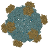
| |||||||||||||||||||||||||||||||||||||||||||||||||||||||||||||||
| 2 |
| |||||||||||||||||||||||||||||||||||||||||||||||||||||||||||||||
| 3 | x 5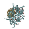
| |||||||||||||||||||||||||||||||||||||||||||||||||||||||||||||||
| 4 | x 6
| |||||||||||||||||||||||||||||||||||||||||||||||||||||||||||||||
| 5 | 
| |||||||||||||||||||||||||||||||||||||||||||||||||||||||||||||||
| 6 | x 20
| |||||||||||||||||||||||||||||||||||||||||||||||||||||||||||||||
| Unit cell |
| |||||||||||||||||||||||||||||||||||||||||||||||||||||||||||||||
| Symmetry | Point symmetry: (Hermann–Mauguin notation: 532 / Schoenflies symbol: I (icosahedral)) | |||||||||||||||||||||||||||||||||||||||||||||||||||||||||||||||
| Noncrystallographic symmetry (NCS) | NCS oper:
|
-Components
| #1: Protein | Mass: 49294.277 Da / Num. of mol.: 1 / Source method: isolated from a natural source / Details: wt alpha3 capsid protein F / Source: (natural) Enterobacteria phage alpha3 (virus) / Genus: Microvirus / References: UniProt: P08767 |
|---|---|
| #2: Protein | Mass: 19598.990 Da / Num. of mol.: 1 / Source method: isolated from a natural source / Details: wt alpha3 spike protein G / Source: (natural) Enterobacteria phage alpha3 (virus) / Genus: Microvirus / References: UniProt: P31281 |
| #3: Protein/peptide | Mass: 4107.821 Da / Num. of mol.: 1 Source method: isolated from a genetically manipulated source Source: (gene. exp.) Enterobacteria phage phiX174 (virus) / Genus: Microvirus / Species: Enterobacteria phage phiX174 sensu lato / Gene: J / Production host:  |
| #4: DNA chain | Mass: 1183.845 Da / Num. of mol.: 1 / Source method: obtained synthetically |
| #5: Chemical | ChemComp-DC /   Type: DNA linking / Mass: 307.197 Da / Num. of mol.: 6 / Source method: obtained synthetically / Formula: C9H14N3O7P / Comment: dCMP*YM Type: DNA linking / Mass: 307.197 Da / Num. of mol.: 6 / Source method: obtained synthetically / Formula: C9H14N3O7P / Comment: dCMP*YM |
-Experimental details
-Experiment
| Experiment | Method: X-RAY DIFFRACTION / Number of used crystals: 1 |
|---|
- Sample preparation
| Crystal grow | Temperature: 298 K / Method: vapor diffusion, sitting drop / pH: 5 Details: 4-7% PEG 8000, 100 mM sodium citrate pH 5.0, 40% glycerol, 0.02% sodium azide, 0.1% beta-mercapto-ethanol, 0.9M NaCl, VAPOR DIFFUSION, SITTING DROP, temperature 298K | ||||||||||||||||||||||||||||||||||||||||||||||||
|---|---|---|---|---|---|---|---|---|---|---|---|---|---|---|---|---|---|---|---|---|---|---|---|---|---|---|---|---|---|---|---|---|---|---|---|---|---|---|---|---|---|---|---|---|---|---|---|---|---|
| Components of the solutions |
|
-Data collection
| Diffraction | Mean temperature: 100 K |
|---|---|
| Diffraction source | Source: SYNCHROTRON / Site: APS  / Beamline: 14-BM-C / Wavelength: 1 Å / Beamline: 14-BM-C / Wavelength: 1 Å |
| Detector | Type: ADSC QUANTUM 4 / Detector: CCD / Date: May 11, 2001 |
| Radiation | Protocol: SINGLE WAVELENGTH / Monochromatic (M) / Laue (L): M / Scattering type: x-ray |
| Radiation wavelength | Wavelength: 1 Å / Relative weight: 1 |
| Reflection | Resolution: 3.5→500 Å / Num. all: 342669 / Num. obs: 269234 / % possible obs: 96.9 % / Observed criterion σ(F): 0 / Observed criterion σ(I): 2 / Biso Wilson estimate: 1 Å2 / Rmerge(I) obs: 0.097 |
| Reflection shell | Resolution: 3.5→3.66 Å / Rmerge(I) obs: 0.157 / % possible all: 94.4 |
- Processing
| Software |
| |||||||||||||||||||||||||
|---|---|---|---|---|---|---|---|---|---|---|---|---|---|---|---|---|---|---|---|---|---|---|---|---|---|---|
| Refinement | Method to determine structure: MOLECULAR REPLACEMENT Starting model: Bacteriophage alpha3 wild-type structure Resolution: 3.5→92.91 Å / Rfactor Rfree error: 0.002 / Data cutoff high absF: 112625.55 / Data cutoff high rms absF: 112625.55 / Data cutoff low absF: 0 / Isotropic thermal model: RESTRAINED / Cross valid method: THROUGHOUT / σ(F): 0 / Stereochemistry target values: Engh & Huber Details: the O3*-P bond between residue (X A 3 ) and Residue (X A 4 ) is 1.91A. The density for the nucleic acid in this region is very weak and therefore difficult to interpret.
| |||||||||||||||||||||||||
| Solvent computation | Solvent model: FLAT MODEL / Bsol: 10 Å2 / ksol: 0.312307 e/Å3 | |||||||||||||||||||||||||
| Displacement parameters | Biso mean: 22.3 Å2
| |||||||||||||||||||||||||
| Refine analyze |
| |||||||||||||||||||||||||
| Refinement step | Cycle: LAST / Resolution: 3.5→92.91 Å
| |||||||||||||||||||||||||
| Refine LS restraints |
| |||||||||||||||||||||||||
| Refine LS restraints NCS | NCS model details: CONSTR | |||||||||||||||||||||||||
| LS refinement shell | Resolution: 3.5→3.66 Å / Rfactor Rfree error: 0.007 / Total num. of bins used: 8
| |||||||||||||||||||||||||
| Xplor file |
|
