 Movie
Movie Controller

[English] 日本語
 Yorodumi
- PDB-2gvc: Crystal structure of flavin-containing monooxygenase (FMO)from S.... -
+ Open data

- Basic information
| Entry | Database: PDB / ID: 2gvc | ||||||
|---|---|---|---|---|---|---|---|
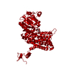
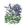
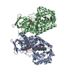
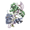
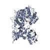
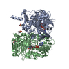
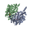
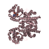
| Title | Crystal structure of flavin-containing monooxygenase (FMO)from S.pombe and substrate (methimazole) complex | ||||||
 Components Components | monooxygenase | ||||||
 Keywords Keywords | OXIDOREDUCTASE / FMO / FAD / methimazole / oxygenase / PSI / Structural Genomics / Protein Structure Initiative / New York SGX Research Center for Structural Genomics / NYSGXRC | ||||||
| Function / homology |  Function and homology information Function and homology informationOxidoreductases; Acting on paired donors, with incorporation or reduction of molecular oxygen; With NADH or NADPH as one donor, and incorporation of one atom of oxygen into the other donor / N,N-dimethylaniline monooxygenase activity / cellular detoxification / FAD binding / monooxygenase activity / NADP binding / endoplasmic reticulum membrane Similarity search - Function | ||||||
| Biological species |  | ||||||
| Method |  X-RAY DIFFRACTION / SYNCHROTRON / MOLECULAR REPLACEMENT / Resolution: 2.22 Å X-RAY DIFFRACTION / SYNCHROTRON / MOLECULAR REPLACEMENT / Resolution: 2.22 Å | ||||||
 Authors Authors | Eswaramoorthy, S. / Swaminathan, S. / Burley, S.K. / New York SGX Research Center for Structural Genomics (NYSGXRC) | ||||||
 Citation Citation | Journal: Proc.Natl.Acad.Sci.Usa / Year: 2006 Title: Mechanism of action of a flavin-containing monooxygenase. Authors: Eswaramoorthy, S. / Bonanno, J.B. / Burley, S.K. / Swaminathan, S. | ||||||
| History |
|
- Structure visualization
| Structure viewer | Molecule: MolmilJmol/JSmol |
|---|
- Downloads & links
-Download
| PDBx/mmCIF format | 2gvc.cif.gz | 368 KB | Display | PDBx/mmCIF format |
|---|---|---|---|---|
| PDB format | pdb2gvc.ent.gz | 299.1 KB | Display | PDB format |
| PDBx/mmJSON format | 2gvc.json.gz | Tree view | PDBx/mmJSON format | |
| Others |  Other downloads Other downloads |
-Validation report
| Arichive directory | https://data.pdbj.org/pub/pdb/validation_reports/gv/2gvcftp://data.pdbj.org/pub/pdb/validation_reports/gv/2gvc | HTTPS FTP |
|---|
-Related structure data
| Related structure data |  1vqwSC  2gv8C S: Starting model for refinement C: citing same article ( |
|---|---|
| Similar structure data | |
| Other databases |
-Links
 PDBj
PDBj- Assembly
| Deposited unit | 
| ||||||||
|---|---|---|---|---|---|---|---|---|---|
| 1 | 
| ||||||||
| 2 | 
| ||||||||
| Unit cell |
|
-Components
-Protein , 1 types, 4 molecules ABDE
| #1: Protein | Mass: 50179.543 Da / Num. of mol.: 4 Source method: isolated from a genetically manipulated source Source: (gene. exp.) Production host:  |
|---|
-Non-polymers , 5 types, 553 molecules 


| #2: Chemical | ChemComp-PEO /  Mass: 34.015 Da / Num. of mol.: 4 / Source method: obtained synthetically / Formula: H2O2 Mass: 34.015 Da / Num. of mol.: 4 / Source method: obtained synthetically / Formula: H2O2#3: Chemical | ChemComp-FAD /  Mass: 785.550 Da / Num. of mol.: 4 / Source method: obtained synthetically / Formula: C27H33N9O15P2 / Comment: FAD*YM Mass: 785.550 Da / Num. of mol.: 4 / Source method: obtained synthetically / Formula: C27H33N9O15P2 / Comment: FAD*YM#4: Chemical | ChemComp-MMZ /  Mass: 114.169 Da / Num. of mol.: 4 / Source method: obtained synthetically / Formula: C4H6N2S / Comment: medication*YM Mass: 114.169 Da / Num. of mol.: 4 / Source method: obtained synthetically / Formula: C4H6N2S / Comment: medication*YM#5: Chemical | ChemComp-GOL /  Mass: 92.094 Da / Num. of mol.: 4 / Source method: obtained synthetically / Formula: C3H8O3 Mass: 92.094 Da / Num. of mol.: 4 / Source method: obtained synthetically / Formula: C3H8O3#6: Water | ChemComp-HOH / | Mass: 18.015 Da / Num. of mol.: 537 / Source method: isolated from a natural source / Formula: H2O |
|---|
-Details
| Has protein modification | Y |
|---|
-Experimental details
-Experiment
| Experiment | Method: X-RAY DIFFRACTION / Number of used crystals: 1 |
|---|
- Sample preparation
| Crystal | Density Matthews: 3.15 Å3/Da / Density % sol: 60.97 % |
|---|---|
| Crystal grow | Temperature: 298 K / Method: vapor diffusion, sitting drop / pH: 5.6 Details: PEG 3350, Ammonium Acetate, pH 5.6, VAPOR DIFFUSION, SITTING DROP, temperature 298K |
-Data collection
| Diffraction | Mean temperature: 100 K |
|---|---|
| Diffraction source | Source: SYNCHROTRON / Site: NSLS  / Beamline: X25 / Wavelength: 1.1 Å / Beamline: X25 / Wavelength: 1.1 Å |
| Detector | Type: ADSC QUANTUM 315 / Detector: CCD / Date: Jul 12, 2005 |
| Radiation | Monochromator: Si 111 CHANNEL / Protocol: SINGLE WAVELENGTH / Monochromatic (M) / Laue (L): M / Scattering type: x-ray |
| Radiation wavelength | Wavelength: 1.1 Å / Relative weight: 1 |
| Reflection | Resolution: 2.22→50 Å / Num. all: 112153 / Num. obs: 112153 / % possible obs: 92.7 % / Observed criterion σ(F): 0 / Observed criterion σ(I): 0 / Redundancy: 3.3 % / Rmerge(I) obs: 0.067 / Net I/σ(I): 9.3 |
| Reflection shell | Resolution: 2.22→2.3 Å / Redundancy: 2.8 % / Rmerge(I) obs: 0.321 / Num. unique all: 9155 / % possible all: 75.7 |
- Processing
| Software |
| ||||||||||||||||||||
|---|---|---|---|---|---|---|---|---|---|---|---|---|---|---|---|---|---|---|---|---|---|
| Refinement | Method to determine structure: MOLECULAR REPLACEMENT Starting model: pdb entry 1VQW Resolution: 2.22→50 Å / Cross valid method: THROUGHOUT / σ(F): 0 / Stereochemistry target values: Engh & Huber Details: The side chain atoms of GLU 444 were not modeled due to lack of electron density.
| ||||||||||||||||||||
| Refinement step | Cycle: LAST / Resolution: 2.22→50 Å
| ||||||||||||||||||||
| Refine LS restraints |
|
