Movie
Movie Controller

[English] 日本語
 Yorodumi
- PDB-7piv: Active Melanocortin-4 receptor (MC4R)- Gs protein complex bound t... -
+ Open data

- Basic information
| Entry | Database: PDB / ID: 7piv | ||||||||||||
|---|---|---|---|---|---|---|---|---|---|---|---|---|---|
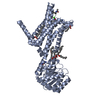
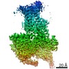
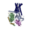
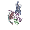
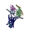
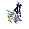
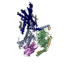
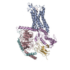
| Title | Active Melanocortin-4 receptor (MC4R)- Gs protein complex bound to agonist NDP-alpha-MSH at 2.86 A resolution. | ||||||||||||
 Components Components |
| ||||||||||||
 Keywords Keywords | SIGNALING PROTEIN / GPCR / MELANOCORTIN-4 RECEPTOR / MELANOCORTIN RECEPTORS / SETMELANOTIDE / NDP-ALPHA-MSH / ALPHA-18 MSH / ANTAGONISM / AGONISM / APPETITE REGULATION / ANTI-OBESITY TREATMENT | ||||||||||||
| Function / homology |  Function and homology information Function and homology informationresponse to melanocyte-stimulating hormone / melanocyte-stimulating hormone receptor activity / G beta:gamma signalling through PLC beta / Presynaptic function of Kainate receptors / Prostacyclin signalling through prostacyclin receptor / G alpha (z) signalling events / Glucagon-type ligand receptors / G beta:gamma signalling through PI3Kgamma / G beta:gamma signalling through CDC42 / melanocortin receptor activity ...response to melanocyte-stimulating hormone / melanocyte-stimulating hormone receptor activity / G beta:gamma signalling through PLC beta / Presynaptic function of Kainate receptors / Prostacyclin signalling through prostacyclin receptor / G alpha (z) signalling events / Glucagon-type ligand receptors / G beta:gamma signalling through PI3Kgamma / G beta:gamma signalling through CDC42 / melanocortin receptor activity / corticotropin receptor activity / non-motile cilium membrane / Adrenaline,noradrenaline inhibits insulin secretion / ADP signalling through P2Y purinoceptor 12 / Cooperation of PDCL (PhLP1) and TRiC/CCT in G-protein beta folding / Thromboxane signalling through TP receptor / G beta:gamma signalling through BTK / Thrombin signalling through proteinase activated receptors (PARs) / Activation of G protein gated Potassium channels / Inhibition of voltage gated Ca2+ channels via Gbeta/gamma subunits / G alpha (s) signalling events / Ca2+ pathway / G-protein activation / regulation of feeding behavior / Extra-nuclear estrogen signaling / G alpha (12/13) signalling events / G alpha (q) signalling events / Vasopressin regulates renal water homeostasis via Aquaporins / GPER1 signaling / Glucagon-like Peptide-1 (GLP1) regulates insulin secretion / ADP signalling through P2Y purinoceptor 1 / High laminar flow shear stress activates signaling by PIEZO1 and PECAM1:CDH5:KDR in endothelial cells / negative regulation of appetite / G alpha (i) signalling events / feeding behavior / positive regulation of bone resorption / G-protein activation / Activation of the phototransduction cascade / Glucagon-type ligand receptors / Thromboxane signalling through TP receptor / Sensory perception of sweet, bitter, and umami (glutamate) taste / G beta:gamma signalling through PI3Kgamma / G beta:gamma signalling through CDC42 / Cooperation of PDCL (PhLP1) and TRiC/CCT in G-protein beta folding / Activation of G protein gated Potassium channels / Inhibition of voltage gated Ca2+ channels via Gbeta/gamma subunits / Ca2+ pathway / G alpha (z) signalling events / High laminar flow shear stress activates signaling by PIEZO1 and PECAM1:CDH5:KDR in endothelial cells / Glucagon-like Peptide-1 (GLP1) regulates insulin secretion / Vasopressin regulates renal water homeostasis via Aquaporins / Adrenaline,noradrenaline inhibits insulin secretion / ADP signalling through P2Y purinoceptor 12 / G alpha (q) signalling events / neuropeptide binding / G alpha (i) signalling events / Thrombin signalling through proteinase activated receptors (PARs) / photoreceptor outer segment membrane / negative regulation of feeding behavior / spectrin binding / sensory perception of taste / alkylglycerophosphoethanolamine phosphodiesterase activity / retina development in camera-type eye / adenylate cyclase-activating G protein-coupled bile acid receptor signaling pathway / adenylate cyclase-activating serotonin receptor signaling pathway / regulation of skeletal muscle contraction / hair follicle placode formation / PKA activation in glucagon signalling / developmental growth / intracellular transport / photoreceptor outer segment / D1 dopamine receptor binding / renal water homeostasis / vascular endothelial cell response to laminar fluid shear stress / Hedgehog 'off' state / activation of adenylate cyclase activity / adenylate cyclase-activating adrenergic receptor signaling pathway / cellular response to acidic pH / cardiac muscle cell apoptotic process / regulation of eating behavior / photoreceptor inner segment / Transcriptional and post-translational regulation of MITF-M expression and activity / cellular response to glucagon stimulus / intracellular glucose homeostasis / adenylate cyclase activator activity / trans-Golgi network membrane / positive regulation of insulin secretion involved in cellular response to glucose stimulus / Peptide ligand-binding receptors / negative regulation of inflammatory response to antigenic stimulus / response to prostaglandin E / bone development / cell population proliferation / platelet aggregation / cognition / positive regulation of insulin secretion / G-protein beta/gamma-subunit complex binding / adenylate cyclase-modulating G protein-coupled receptor signaling pathway / phospholipase C-activating G protein-coupled receptor signaling pathway / sensory perception of smell / Glucagon signaling in metabolic regulation Similarity search - Function | ||||||||||||
| Biological species |  Homo sapiens (human) Homo sapiens (human) synthetic construct (others) | ||||||||||||
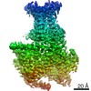
| Method | ELECTRON MICROSCOPY / single particle reconstruction / cryo EM / Resolution: 2.86 Å | ||||||||||||
 Authors Authors | Heyder, N.A. / Schmidt, A. / Kleinau, G. / Hilal, T. / Scheerer, P. | ||||||||||||
| Funding support |  Germany, 3items Germany, 3items
| ||||||||||||
 Citation Citation | Journal: Cell Res / Year: 2021 Title: Structures of active melanocortin-4 receptor-Gs-protein complexes with NDP-α-MSH and setmelanotide. Authors: Nicolas A Heyder / Gunnar Kleinau / David Speck / Andrea Schmidt / Sarah Zschunke / Michal Szczepek / Brian Bauer / Anja Koch / Monique Gallandi / Dennis Kwiatkowski / Jörg Bürger / ...Authors: Nicolas A Heyder / Gunnar Kleinau / David Speck / Andrea Schmidt / Sarah Zschunke / Michal Szczepek / Brian Bauer / Anja Koch / Monique Gallandi / Dennis Kwiatkowski / Jörg Bürger / Thorsten Mielke / Annette G Beck-Sickinger / Peter W Hildebrand / Christian M T Spahn / Daniel Hilger / Magdalena Schacherl / Heike Biebermann / Tarek Hilal / Peter Kühnen / Brian K Kobilka / Patrick Scheerer /  Abstract: The melanocortin-4 receptor (MC4R), a hypothalamic master regulator of energy homeostasis and appetite, is a class A G-protein-coupled receptor and a prime target for the pharmacological treatment of ...The melanocortin-4 receptor (MC4R), a hypothalamic master regulator of energy homeostasis and appetite, is a class A G-protein-coupled receptor and a prime target for the pharmacological treatment of obesity. Here, we present cryo-electron microscopy structures of MC4R-Gs-protein complexes with two drugs recently approved by the FDA, the peptide agonists NDP-α-MSH and setmelanotide, with 2.9 Å and 2.6 Å resolution. Together with signaling data from structure-derived MC4R mutants, the complex structures reveal the agonist-induced origin of transmembrane helix (TM) 6-regulated receptor activation. The ligand-binding modes of NDP-α-MSH, a high-affinity linear variant of the endogenous agonist α-MSH, and setmelanotide, a cyclic anti-obesity drug with biased signaling toward Gq/11, underline the key role of TM3 in ligand-specific interactions and of calcium ion as a ligand-adaptable cofactor. The agonist-specific TM3 interplay subsequently impacts receptor-Gs-protein interfaces at intracellular loop 2, which also regulates the G-protein coupling profile of this promiscuous receptor. Finally, our structures reveal mechanistic details of MC4R activation/inhibition, and provide important insights into the regulation of the receptor signaling profile which will facilitate the development of tailored anti-obesity drugs. | ||||||||||||
| History |
|
- Structure visualization
| Movie |
Movie viewer |
|---|---|
| Structure viewer | Molecule: MolmilJmol/JSmol |
- Downloads & links
-Download
| PDBx/mmCIF format | 7piv.cif.gz | 217.2 KB | Display | PDBx/mmCIF format |
|---|---|---|---|---|
| PDB format | pdb7piv.ent.gz | 163.9 KB | Display | PDB format |
| PDBx/mmJSON format | 7piv.json.gz | Tree view | PDBx/mmJSON format | |
| Others |  Other downloads Other downloads |
-Validation report
| Arichive directory | https://data.pdbj.org/pub/pdb/validation_reports/pi/7pivftp://data.pdbj.org/pub/pdb/validation_reports/pi/7piv | HTTPS FTP |
|---|
-Related structure data
| Related structure data | 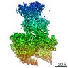 13454MC 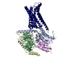 7piuC M: map data used to model this data C: citing same article ( |
|---|---|
| Similar structure data |


-Links
 PDBj
PDBj


- Assembly
| Deposited unit | 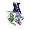
|
|---|---|
| 1 |
|
-Components
-Protein , 2 types, 2 molecules RA
| #1: Protein | Mass: 38698.520 Da / Num. of mol.: 1 Source method: isolated from a genetically manipulated source Source: (gene. exp.) Homo sapiens (human) / Gene: MC4R / Plasmid: pOET3 / Production host:   Spodoptera frugiperda (fall armyworm) / References: UniProt: P32245 Spodoptera frugiperda (fall armyworm) / References: UniProt: P32245 |
|---|---|
| #3: Protein | Mass: 44326.160 Da / Num. of mol.: 1 Source method: isolated from a genetically manipulated source Source: (gene. exp.) Homo sapiens (human) / Gene: GNAS, GNAS1, GSP / Variant: 2 / Plasmid: pFastbac / Production host: Trichoplusia ni (cabbage looper) / References: UniProt: P63092 |
-Guanine nucleotide-binding protein ... , 2 types, 2 molecules BG
| #4: Protein | Mass: 37728.152 Da / Num. of mol.: 1 Source method: isolated from a genetically manipulated source Source: (gene. exp.) Trichoplusia ni (cabbage looper) / References: UniProt: P54311 |
|---|---|
| #5: Protein | Mass: 7861.143 Da / Num. of mol.: 1 Source method: isolated from a genetically manipulated source Source: (gene. exp.) Trichoplusia ni (cabbage looper) / References: UniProt: P63213 |
-Protein/peptide / Antibody , 2 types, 2 molecules PN
| #2: Protein/peptide | Mass: 1632.863 Da / Num. of mol.: 1 / Source method: obtained synthetically / Source: (synth.) synthetic construct (others) |
|---|---|
| #6: Antibody | Mass: 14714.320 Da / Num. of mol.: 1 Source method: isolated from a genetically manipulated source Source: (gene. exp.)  |
-Non-polymers , 2 types, 116 molecules 

| #7: Chemical | ChemComp-CA /  Mass: 40.078 Da / Num. of mol.: 1 / Source method: obtained synthetically / Formula: Ca / Feature type: SUBJECT OF INVESTIGATION Mass: 40.078 Da / Num. of mol.: 1 / Source method: obtained synthetically / Formula: Ca / Feature type: SUBJECT OF INVESTIGATION |
|---|---|
| #8: Water | ChemComp-HOH / Mass: 18.015 Da / Num. of mol.: 115 / Source method: isolated from a natural source / Formula: H2O |
-Details
| Has ligand of interest | Y |
|---|---|
| Has protein modification | Y |
-Experimental details
-Experiment
| Experiment | Method: ELECTRON MICROSCOPY |
|---|---|
| EM experiment | Aggregation state: PARTICLE / 3D reconstruction method: single particle reconstruction |
- Sample preparation
| Component | Name: Ternary complex of the NDP-alpha-MSH activated melanocortin 4 receptor (MC4R) Gs protein complex stabilized by nanobody 35 Type: COMPLEX / Entity ID: #1-#6 / Source: MULTIPLE SOURCES | ||||||||||||||||||||||||||||||||||||||||
|---|---|---|---|---|---|---|---|---|---|---|---|---|---|---|---|---|---|---|---|---|---|---|---|---|---|---|---|---|---|---|---|---|---|---|---|---|---|---|---|---|---|
| Molecular weight | Value: 144.6 kDa/nm / Experimental value: NO | ||||||||||||||||||||||||||||||||||||||||
| Source (natural) |
| ||||||||||||||||||||||||||||||||||||||||
| Buffer solution | pH: 7.5 | ||||||||||||||||||||||||||||||||||||||||
| Buffer component |
| ||||||||||||||||||||||||||||||||||||||||
| Specimen | Conc.: 1.2 mg/ml / Embedding applied: NO / Shadowing applied: NO / Staining applied: NO / Vitrification applied: YES | ||||||||||||||||||||||||||||||||||||||||
| Specimen support | Grid material: GOLD / Grid type: UltrAuFoil R1.2/1.3 | ||||||||||||||||||||||||||||||||||||||||
| Vitrification | Instrument: FEI VITROBOT MARK IV / Cryogen name: ETHANE / Humidity: 100 % / Chamber temperature: 283 K |
- Electron microscopy imaging
| Experimental equipment |  Model: Titan Krios / Image courtesy: FEI Company |
|---|---|
| Microscopy | Model: FEI TITAN KRIOS |
| Electron gun | Electron source:  FIELD EMISSION GUN / Accelerating voltage: 300 kV / Illumination mode: FLOOD BEAM FIELD EMISSION GUN / Accelerating voltage: 300 kV / Illumination mode: FLOOD BEAM |
| Electron lens | Mode: BRIGHT FIELD / Nominal magnification: 96000 X / Nominal defocus max: 2000 nm / Nominal defocus min: 800 nm / C2 aperture diameter: 50 µm / Alignment procedure: ZEMLIN TABLEAU |
| Specimen holder | Cryogen: NITROGEN / Specimen holder model: FEI TITAN KRIOS AUTOGRID HOLDER |
| Image recording | Average exposure time: 40.78 sec. / Electron dose: 40 e/Å2 / Detector mode: COUNTING / Film or detector model: FEI FALCON III (4k x 4k) / Num. of grids imaged: 1 / Num. of real images: 5618 |
- Processing
| EM software |
| ||||||||||||||||||||||||||||||||||||||||||||||||
|---|---|---|---|---|---|---|---|---|---|---|---|---|---|---|---|---|---|---|---|---|---|---|---|---|---|---|---|---|---|---|---|---|---|---|---|---|---|---|---|---|---|---|---|---|---|---|---|---|---|
| CTF correction | Type: PHASE FLIPPING AND AMPLITUDE CORRECTION | ||||||||||||||||||||||||||||||||||||||||||||||||
| Particle selection | Num. of particles selected: 2746119 | ||||||||||||||||||||||||||||||||||||||||||||||||
| Symmetry | Point symmetry: C1 (asymmetric) | ||||||||||||||||||||||||||||||||||||||||||||||||
| 3D reconstruction | Resolution: 2.86 Å / Resolution method: FSC 0.143 CUT-OFF / Num. of particles: 221682 / Symmetry type: POINT | ||||||||||||||||||||||||||||||||||||||||||||||||
| Atomic model building | Protocol: OTHER / Space: REAL | ||||||||||||||||||||||||||||||||||||||||||||||||
| Atomic model building |
|