 Movie
Movie Controller

[English] 日本語
 Yorodumi
- PDB-7o4c: Crystal structure of PASTA domains of the Penicillin-Binding Prot... -
+ Open data

- Basic information
| Entry | Database: PDB / ID: 7o4c | ||||||
|---|---|---|---|---|---|---|---|
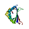
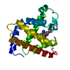
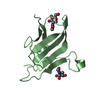
| Title | Crystal structure of PASTA domains of the Penicillin-Binding Protein 1 (PBP1) from Staphylococcus aureus | ||||||
 Components Components | Penicillin-binding protein 1 | ||||||
 Keywords Keywords | HYDROLASE / Cell division / antibiotic resistance / peptidoglycan synthesis / transpeptidase / penicillin-binding protein | ||||||
| Function / homology |  Function and homology information Function and homology information | ||||||
| Biological species |  Staphylococcus aureus subsp. aureus COL (bacteria) Staphylococcus aureus subsp. aureus COL (bacteria) | ||||||
| Method |  X-RAY DIFFRACTION / SYNCHROTRON / MOLECULAR REPLACEMENT / Resolution: 1.51 Å X-RAY DIFFRACTION / SYNCHROTRON / MOLECULAR REPLACEMENT / Resolution: 1.51 Å | ||||||
 Authors Authors | Martinez Caballero, S. / Hermoso, J.A. | ||||||
 Citation Citation | Journal: Comput Struct Biotechnol J / Year: 2021 Title: Integrative structural biology of the penicillin-binding protein-1 from Staphylococcus aureus , an essential component of the divisome machinery. Authors: Martinez-Caballero, S. / Mahasenan, K.V. / Kim, C. / Molina, R. / Feltzer, R. / Lee, M. / Bouley, R. / Hesek, D. / Fisher, J.F. / Munoz, I.G. / Chang, M. / Mobashery, S. / Hermoso, J.A. | ||||||
| History |
|
- Structure visualization
| Structure viewer | Molecule: MolmilJmol/JSmol |
|---|
- Downloads & links
-Download
| PDBx/mmCIF format | 7o4c.cif.gz | 113.5 KB | Display | PDBx/mmCIF format |
|---|---|---|---|---|
| PDB format | pdb7o4c.ent.gz | 86.8 KB | Display | PDB format |
| PDBx/mmJSON format | 7o4c.json.gz | Tree view | PDBx/mmJSON format | |
| Others |  Other downloads Other downloads |
-Validation report
| Arichive directory | https://data.pdbj.org/pub/pdb/validation_reports/o4/7o4cftp://data.pdbj.org/pub/pdb/validation_reports/o4/7o4c | HTTPS FTP |
|---|
-Related structure data
| Related structure data |  7o49C  7o4aC  7o4bC  7ok9C 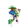 5oauS S: Starting model for refinement C: citing same article ( |
|---|---|
| Similar structure data |


-Links
 PDBj
PDBj
- Assembly
| Deposited unit | 
| ||||||||
|---|---|---|---|---|---|---|---|---|---|
| 1 | 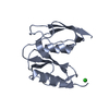
| ||||||||
| 2 | 
| ||||||||
| Unit cell |
|
-Components
| #1: Protein | Mass: 12647.298 Da / Num. of mol.: 2 Source method: isolated from a genetically manipulated source Source: (gene. exp.) Staphylococcus aureus subsp. aureus COL (bacteria)Gene: pbp1, SACOL1194 / Production host: #2: Chemical | ChemComp-CL / |   Mass: 35.453 Da / Num. of mol.: 1 / Source method: isolated from a natural source / Formula: Cl Mass: 35.453 Da / Num. of mol.: 1 / Source method: isolated from a natural source / Formula: Cl#3: Water | ChemComp-HOH / |  Mass: 18.015 Da / Num. of mol.: 166 / Source method: isolated from a natural source / Formula: H2O Mass: 18.015 Da / Num. of mol.: 166 / Source method: isolated from a natural source / Formula: H2OHas ligand of interest | N | |
|---|
-Experimental details
-Experiment
| Experiment | Method: X-RAY DIFFRACTION / Number of used crystals: 1 |
|---|
- Sample preparation
| Crystal | Density Matthews: 2.88 Å3/Da / Density % sol: 57.26 % |
|---|---|
| Crystal grow | Temperature: 291.15 K / Method: vapor diffusion, sitting drop Details: PEG 400 HEPES free acid/sodium hydroxide PEG 3000 Glycerol |
-Data collection
| Diffraction | Mean temperature: 100 K / Serial crystal experiment: N |
|---|---|
| Diffraction source | Source: SYNCHROTRON / Site: ALBA  / Beamline: XALOC / Wavelength: 0.97918 Å / Beamline: XALOC / Wavelength: 0.97918 Å |
| Detector | Type: DECTRIS PILATUS 6M / Detector: PIXEL / Date: Jul 11, 2020 |
| Radiation | Protocol: SINGLE WAVELENGTH / Monochromatic (M) / Laue (L): M / Scattering type: x-ray |
| Radiation wavelength | Wavelength: 0.97918 Å / Relative weight: 1 |
| Reflection | Resolution: 1.51→42.51 Å / Num. obs: 46753 / % possible obs: 100 % / Redundancy: 13 % / Rmerge(I) obs: 0.042 / Rpim(I) all: 0.012 / Net I/σ(I): 30.8 |
| Reflection shell | Resolution: 1.51→1.54 Å / Rmerge(I) obs: 0.687 / Num. unique obs: 2309 / Rpim(I) all: 0.193 |
- Processing
| Software |
| ||||||||||||||||||||||||||||||||||||||||||||||||||||||||||||
|---|---|---|---|---|---|---|---|---|---|---|---|---|---|---|---|---|---|---|---|---|---|---|---|---|---|---|---|---|---|---|---|---|---|---|---|---|---|---|---|---|---|---|---|---|---|---|---|---|---|---|---|---|---|---|---|---|---|---|---|---|---|
| Refinement | Method to determine structure: MOLECULAR REPLACEMENT Starting model: 5OAU Resolution: 1.51→40.31 Å / Cor.coef. Fo:Fc: 0.974 / Cor.coef. Fo:Fc free: 0.959 / SU B: 2.456 / SU ML: 0.041 / Cross valid method: THROUGHOUT / σ(F): 0 / ESU R: 0.07 / ESU R Free: 0.069 / Stereochemistry target values: MAXIMUM LIKELIHOOD Details: HYDROGENS HAVE BEEN ADDED IN THE RIDING POSITIONS U VALUES : REFINED INDIVIDUALLY
| ||||||||||||||||||||||||||||||||||||||||||||||||||||||||||||
| Solvent computation | Ion probe radii: 0.8 Å / Shrinkage radii: 0.8 Å / VDW probe radii: 1.2 Å / Solvent model: MASK | ||||||||||||||||||||||||||||||||||||||||||||||||||||||||||||
| Displacement parameters | Biso max: 169.98 Å2 / Biso mean: 31.602 Å2 / Biso min: 14.79 Å2
| ||||||||||||||||||||||||||||||||||||||||||||||||||||||||||||
| Refinement step | Cycle: final / Resolution: 1.51→40.31 Å
| ||||||||||||||||||||||||||||||||||||||||||||||||||||||||||||
| Refine LS restraints |
| ||||||||||||||||||||||||||||||||||||||||||||||||||||||||||||
| LS refinement shell | Resolution: 1.51→1.549 Å / Rfactor Rfree error: 0 / Total num. of bins used: 20
|
