Movie
Movie Controller


+ Open data

- Basic information
| Entry | Database: PDB / ID: 7o3h | ||||||
|---|---|---|---|---|---|---|---|
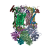
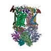
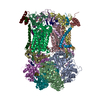
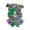
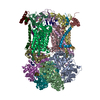
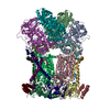
| Title | Murine CIII2 focus-refined from supercomplex CICIII2 | ||||||
 Components Components |
| ||||||
 Keywords Keywords | MEMBRANE PROTEIN / mitochondria / respiratory chain / supercomplex / OXIDOREDUCTASE / complex III / complex IV | ||||||
| Function / homology |  Function and homology information Function and homology informationresponse to D-galactosamine / Complex III assembly / response to cobalamin / Mitochondrial translation termination / Respiratory electron transport / response to mercury ion / subthalamus development / pons development / cerebellar Purkinje cell layer development / mitochondrial respiratory chain complex III assembly ...response to D-galactosamine / Complex III assembly / response to cobalamin / Mitochondrial translation termination / Respiratory electron transport / response to mercury ion / subthalamus development / pons development / cerebellar Purkinje cell layer development / mitochondrial respiratory chain complex III assembly / thalamus development / Mitochondrial protein degradation / pyramidal neuron development / response to alkaloid / response to glucagon / respiratory chain complex III / cellular respiration / quinol-cytochrome-c reductase / quinol-cytochrome-c reductase activity / midbrain development / mitochondrial electron transport, ubiquinol to cytochrome c / response to copper ion / hypothalamus development / animal organ regeneration / electron transport coupled proton transport / response to hyperoxia / response to cadmium ion / response to hormone / response to activity / hippocampus development / respiratory electron transport chain / response to calcium ion / metalloendopeptidase activity / 2 iron, 2 sulfur cluster binding / response to toxic substance / mitochondrial membrane / myelin sheath / response to ethanol / response to hypoxia / oxidoreductase activity / mitochondrial inner membrane / response to xenobiotic stimulus / heme binding / ubiquitin protein ligase binding / protein-containing complex binding / protein-containing complex / mitochondrion / proteolysis / nucleoplasm / membrane / metal ion binding Similarity search - Function | ||||||
| Biological species |  | ||||||
| Method | ELECTRON MICROSCOPY / single particle reconstruction / cryo EM / Resolution: 2.6 Å | ||||||
 Authors Authors | Vercellino, I. / Sazanov, L.A. | ||||||
| Funding support |  Austria, 1items Austria, 1items
| ||||||
 Citation Citation | Journal: Nature / Year: 2021 Title: Structure and assembly of the mammalian mitochondrial supercomplex CIIICIV. Authors: Irene Vercellino / Leonid A Sazanov / Abstract: The enzymes of the mitochondrial electron transport chain are key players of cell metabolism. Despite being active when isolated, in vivo they associate into supercomplexes, whose precise role is ...The enzymes of the mitochondrial electron transport chain are key players of cell metabolism. Despite being active when isolated, in vivo they associate into supercomplexes, whose precise role is debated. Supercomplexes CIIICIV (refs. ), CICIII (ref. ) and CICIIICIV (respirasome) exist in mammals, but in contrast to CICIII and the respirasome, to date the only known eukaryotic structures of CIIICIV come from Saccharomyces cerevisiae and plants, which have different organization. Here we present the first, to our knowledge, structures of mammalian (mouse and ovine) CIIICIV and its assembly intermediates, in different conformations. We describe the assembly of CIIICIV from the CIII precursor to the final CIIICIV conformation, driven by the insertion of the N terminus of the assembly factor SCAF1 (ref. ) deep into CIII, while its C terminus is integrated into CIV. Our structures (which include CICIII and the respirasome) also confirm that SCAF1 is exclusively required for the assembly of CIIICIV and has no role in the assembly of the respirasome. We show that CIII is asymmetric due to the presence of only one copy of subunit 9, which straddles both monomers and prevents the attachment of a second copy of SCAF1 to CIII, explaining the presence of one copy of CIV in CIIICIV in mammals. Finally, we show that CIII and CIV gain catalytic advantage when assembled into the supercomplex and propose a role for CIIICIV in fine tuning the efficiency of electron transfer in the electron transport chain. | ||||||
| History |
|
- Structure visualization
| Movie |
Movie viewer |
|---|---|
| Structure viewer | Molecule: MolmilJmol/JSmol |
- Downloads & links
-Download
| PDBx/mmCIF format | 7o3h.cif.gz | 980.2 KB | Display | PDBx/mmCIF format |
|---|---|---|---|---|
| PDB format | pdb7o3h.ent.gz | 683.6 KB | Display | PDB format |
| PDBx/mmJSON format | 7o3h.json.gz | Tree view | PDBx/mmJSON format | |
| Others |  Other downloads Other downloads |
-Validation report
| Arichive directory | https://data.pdbj.org/pub/pdb/validation_reports/o3/7o3hftp://data.pdbj.org/pub/pdb/validation_reports/o3/7o3h | HTTPS FTP |
|---|
-Related structure data
| Related structure data |  12706MC  7o37C  7o3cC  7o3eC C: citing same article ( M: map data used to model this data |
|---|---|
| Similar structure data |


-Links
 PDBj
PDBj


- Assembly
| Deposited unit | 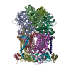
|
|---|---|
| 1 |
|
-Components
-Cytochrome b-c1 complex subunit ... , 9 types, 17 molecules ALBMEPFQGRHSJUKVT
| #1: Protein | Mass: 49355.301 Da / Num. of mol.: 2 / Source method: isolated from a natural source / Source: (natural) #2: Protein | Mass: 46641.773 Da / Num. of mol.: 2 / Source method: isolated from a natural source / Source: (natural) #5: Protein | Mass: 21524.398 Da / Num. of mol.: 2 / Source method: isolated from a natural source / Source: (natural) #6: Protein | Mass: 13456.336 Da / Num. of mol.: 2 / Source method: isolated from a natural source / Source: (natural) #7: Protein | Mass: 9652.051 Da / Num. of mol.: 2 / Source method: isolated from a natural source / Source: (natural) #8: Protein | Mass: 9002.015 Da / Num. of mol.: 2 / Source method: isolated from a natural source / Source: (natural) #9: Protein | Mass: 7326.330 Da / Num. of mol.: 2 / Source method: isolated from a natural source / Source: (natural) #10: Protein | Mass: 6546.627 Da / Num. of mol.: 2 / Source method: isolated from a natural source / Source: (natural) #11: Protein | | Mass: 7900.234 Da / Num. of mol.: 1 / Source method: isolated from a natural source / Source: (natural) |
|---|
-Protein , 2 types, 4 molecules CNDO
| #3: Protein | Mass: 43240.305 Da / Num. of mol.: 2 / Source method: isolated from a natural source / Source: (natural) #4: Protein | Mass: 27312.188 Da / Num. of mol.: 2 / Source method: isolated from a natural source / Source: (natural) |
|---|
-Non-polymers , 6 types, 24 molecules 


| #12: Chemical | ChemComp-3PE /  Mass: 748.065 Da / Num. of mol.: 8 / Source method: obtained synthetically / Formula: C41H82NO8P / Comment: phospholipid*YM Mass: 748.065 Da / Num. of mol.: 8 / Source method: obtained synthetically / Formula: C41H82NO8P / Comment: phospholipid*YM#13: Chemical | ChemComp-CDL /  Mass: 1464.043 Da / Num. of mol.: 6 / Source method: obtained synthetically / Formula: C81H156O17P2 / Comment: phospholipid*YM Mass: 1464.043 Da / Num. of mol.: 6 / Source method: obtained synthetically / Formula: C81H156O17P2 / Comment: phospholipid*YM#14: Chemical | ChemComp-HEM /  Mass: 616.487 Da / Num. of mol.: 4 / Source method: obtained synthetically / Formula: C34H32FeN4O4 Mass: 616.487 Da / Num. of mol.: 4 / Source method: obtained synthetically / Formula: C34H32FeN4O4#15: Chemical |  Mass: 618.503 Da / Num. of mol.: 2 / Source method: obtained synthetically / Formula: C34H34FeN4O4 Mass: 618.503 Da / Num. of mol.: 2 / Source method: obtained synthetically / Formula: C34H34FeN4O4#16: Chemical |  Mass: 175.820 Da / Num. of mol.: 2 / Source method: obtained synthetically / Formula: Fe2S2 Mass: 175.820 Da / Num. of mol.: 2 / Source method: obtained synthetically / Formula: Fe2S2#17: Chemical |  Mass: 790.145 Da / Num. of mol.: 2 / Source method: obtained synthetically / Formula: C44H88NO8P / Comment: phospholipid*YM Mass: 790.145 Da / Num. of mol.: 2 / Source method: obtained synthetically / Formula: C44H88NO8P / Comment: phospholipid*YM |
|---|
-Details
| Has ligand of interest | N |
|---|---|
| Has protein modification | Y |
-Experimental details
-Experiment
| Experiment | Method: ELECTRON MICROSCOPY |
|---|---|
| EM experiment | Aggregation state: PARTICLE / 3D reconstruction method: single particle reconstruction |
- Sample preparation
| Component | Name: Murine complex CIII2 focus-refined from supercomplex CICIII2 Type: COMPLEX / Details: Dimer of Complex III / Entity ID: #1-#11 / Source: NATURAL | |||||||||||||||||||||||||
|---|---|---|---|---|---|---|---|---|---|---|---|---|---|---|---|---|---|---|---|---|---|---|---|---|---|---|
| Molecular weight | Value: 0.5 MDa / Experimental value: YES | |||||||||||||||||||||||||
| Source (natural) | Organism: | |||||||||||||||||||||||||
| Buffer solution | pH: 7.7 / Details: CHAPS was added only upon freezing | |||||||||||||||||||||||||
| Buffer component |
| |||||||||||||||||||||||||
| Specimen | Conc.: 4 mg/ml / Embedding applied: NO / Shadowing applied: NO / Staining applied: NO / Vitrification applied: YES | |||||||||||||||||||||||||
| Specimen support | Grid material: COPPER / Grid mesh size: 300 divisions/in. / Grid type: Quantifoil R0.6/1 | |||||||||||||||||||||||||
| Vitrification | Instrument: FEI VITROBOT MARK IV / Cryogen name: ETHANE / Humidity: 100 % / Chamber temperature: 277 K |
- Electron microscopy imaging
| Experimental equipment |  Model: Titan Krios / Image courtesy: FEI Company |
|---|---|
| Microscopy | Model: FEI TITAN KRIOS |
| Electron gun | Electron source:  FIELD EMISSION GUN / Accelerating voltage: 300 kV / Illumination mode: FLOOD BEAM FIELD EMISSION GUN / Accelerating voltage: 300 kV / Illumination mode: FLOOD BEAM |
| Electron lens | Mode: BRIGHT FIELD / Nominal magnification: 75000 X / Nominal defocus max: 1600 nm / Nominal defocus min: 400 nm / Calibrated defocus min: 200 nm / Calibrated defocus max: 2700 nm / Cs: 2.7 mm / C2 aperture diameter: 50 µm / Alignment procedure: COMA FREE |
| Specimen holder | Cryogen: NITROGEN / Specimen holder model: FEI TITAN KRIOS AUTOGRID HOLDER |
| Image recording | Average exposure time: 1.17 sec. / Electron dose: 90.66 e/Å2 / Detector mode: INTEGRATING / Film or detector model: FEI FALCON III (4k x 4k) / Num. of grids imaged: 1 / Num. of real images: 7245 |
- Processing
| Software |
| |||||||||||||||||||||||||||||||||||||||||||||||||||||||||||||||||
|---|---|---|---|---|---|---|---|---|---|---|---|---|---|---|---|---|---|---|---|---|---|---|---|---|---|---|---|---|---|---|---|---|---|---|---|---|---|---|---|---|---|---|---|---|---|---|---|---|---|---|---|---|---|---|---|---|---|---|---|---|---|---|---|---|---|---|
| EM software |
| |||||||||||||||||||||||||||||||||||||||||||||||||||||||||||||||||
| CTF correction | Type: PHASE FLIPPING AND AMPLITUDE CORRECTION | |||||||||||||||||||||||||||||||||||||||||||||||||||||||||||||||||
| Particle selection | Num. of particles selected: 1500000 Details: multiple rounds of picking and classification performed, first with Relion LoG and then with Topaz, to extract the best particles from the non-pure starting material | |||||||||||||||||||||||||||||||||||||||||||||||||||||||||||||||||
| Symmetry | Point symmetry: C1 (asymmetric) | |||||||||||||||||||||||||||||||||||||||||||||||||||||||||||||||||
| 3D reconstruction | Resolution: 2.6 Å / Resolution method: FSC 0.143 CUT-OFF / Num. of particles: 129 / Algorithm: FOURIER SPACE Details: the final pool of particles was classified without alignment around the MPP cavity to separate the two orientations of Sub 9. the two classes were then refined, first globally and then ...Details: the final pool of particles was classified without alignment around the MPP cavity to separate the two orientations of Sub 9. the two classes were then refined, first globally and then focused on CIII, re-oriented along the C2 symmetry axis, to allow for subsequent 180 degrees flipping of one class and final re-pooled refinement with all particles having the same orientation of Sub 9 Num. of class averages: 1 / Symmetry type: POINT | |||||||||||||||||||||||||||||||||||||||||||||||||||||||||||||||||
| Atomic model building | B value: 65 / Protocol: OTHER / Space: REAL / Target criteria: MAXIMAL LIKELIHOOD | |||||||||||||||||||||||||||||||||||||||||||||||||||||||||||||||||
| Atomic model building |
| |||||||||||||||||||||||||||||||||||||||||||||||||||||||||||||||||
| Refinement | Cross valid method: NONE Stereochemistry target values: GeoStd + Monomer Library + CDL v1.2 | |||||||||||||||||||||||||||||||||||||||||||||||||||||||||||||||||
| Displacement parameters | Biso mean: 65.71 Å2 | |||||||||||||||||||||||||||||||||||||||||||||||||||||||||||||||||
| Refine LS restraints |
|