 Movie
Movie Controller

[English] 日本語
 Yorodumi
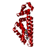
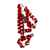
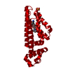
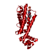
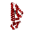
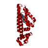
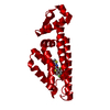
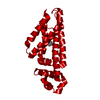
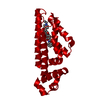
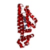
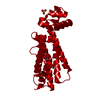
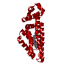
- PDB-7ngt: Mycobacterium tuberculosis transcriptional regulator EthR with bo... -
+ Open data

- Basic information
| Entry | Database: PDB / ID: 7ngt | ||||||
|---|---|---|---|---|---|---|---|
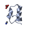
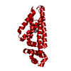
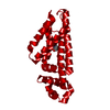
| Title | Mycobacterium tuberculosis transcriptional regulator EthR with bound inhibitory compound | ||||||
 Components Components | HTH-type transcriptional regulator EthR | ||||||
 Keywords Keywords | TRANSCRIPTION / ETHR / Tuberculosis / Inhibition | ||||||
| Function / homology |  Function and homology information Function and homology informationtranscription cis-regulatory region binding / DNA-binding transcription factor activity / response to antibiotic / negative regulation of DNA-templated transcription / regulation of DNA-templated transcription / cytosol Similarity search - Function | ||||||
| Biological species |   Mycobacterium tuberculosis (bacteria) Mycobacterium tuberculosis (bacteria) | ||||||
| Method |  X-RAY DIFFRACTION / SYNCHROTRON / MOLECULAR REPLACEMENT / Resolution: 2.49 Å X-RAY DIFFRACTION / SYNCHROTRON / MOLECULAR REPLACEMENT / Resolution: 2.49 Å | ||||||
 Authors Authors | Tomlinson, C.W.E. / Tatum, N.J. / Pohl, E. | ||||||
 Citation Citation | Journal: To Be Published Title: Systematic exploration of the hydrophobic capacity of the EthR binding site for lead compound optimization Authors: Tatum, N.J. / Tomlinson, C.W.E. / Frita, R. / Bennett, R. / Baulard, A.R. / Pohl, E. / Kitching, M.O. | ||||||
| History |
|
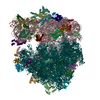
- Structure visualization
| Structure viewer | Molecule: MolmilJmol/JSmol |
|---|
- Downloads & links
-Download
| PDBx/mmCIF format | 7ngt.cif.gz | 52.2 KB | Display | PDBx/mmCIF format |
|---|---|---|---|---|
| PDB format | pdb7ngt.ent.gz | 36.6 KB | Display | PDB format |
| PDBx/mmJSON format | 7ngt.json.gz | Tree view | PDBx/mmJSON format | |
| Others |  Other downloads Other downloads |
-Validation report
| Summary document | 7ngt_validation.pdf.gz | 654.7 KB | Display | wwPDB validaton report |
|---|---|---|---|---|
| Full document | 7ngt_full_validation.pdf.gz | 657.6 KB | Display | |
| Data in XML | 7ngt_validation.xml.gz | 9.4 KB | Display | |
| Data in CIF | 7ngt_validation.cif.gz | 11.9 KB | Display | |
| Arichive directory | https://data.pdbj.org/pub/pdb/validation_reports/ng/7ngtftp://data.pdbj.org/pub/pdb/validation_reports/ng/7ngt | HTTPS FTP |
-Related structure data
| Related structure data | 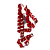 7ngdC 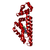 7nggC  7ngiC 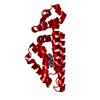 7ngjC 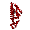 7ngkC 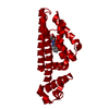 7ngmC 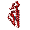 7ngnC 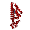 7ngoC 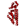 7ngrC  7ngsC 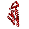 7nguC 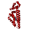 7ngwC 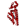 7ngxC 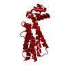 7ngyC 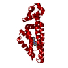 5nioS S: Starting model for refinement C: citing same article ( |
|---|---|
| Similar structure data |


-Links
 PDBj
PDBj- Assembly
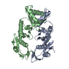
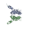
| Deposited unit | 
| ||||||||
|---|---|---|---|---|---|---|---|---|---|
| 1 | 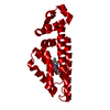
| ||||||||
| Unit cell |
| ||||||||
| Components on special symmetry positions |
|
-Components
| #1: Protein | Mass: 23781.705 Da / Num. of mol.: 1 Source method: isolated from a genetically manipulated source Source: (gene. exp.) Mycobacterium tuberculosis (bacteria) / Gene: ethR, etaR, Rv3855 / Production host: |
|---|---|
| #2: Chemical | ChemComp-8YH / ~{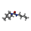  Mass: 198.305 Da / Num. of mol.: 1 / Source method: obtained synthetically / Formula: C11H22N2O / Feature type: SUBJECT OF INVESTIGATION Mass: 198.305 Da / Num. of mol.: 1 / Source method: obtained synthetically / Formula: C11H22N2O / Feature type: SUBJECT OF INVESTIGATION |
| #3: Chemical | ChemComp-SO4 /   Mass: 96.063 Da / Num. of mol.: 1 / Source method: obtained synthetically / Formula: SO4 Mass: 96.063 Da / Num. of mol.: 1 / Source method: obtained synthetically / Formula: SO4 |
| #4: Water | ChemComp-HOH /  Mass: 18.015 Da / Num. of mol.: 5 / Source method: isolated from a natural source / Formula: H2O Mass: 18.015 Da / Num. of mol.: 5 / Source method: isolated from a natural source / Formula: H2O |
| Has ligand of interest | Y |
-Experimental details
-Experiment
| Experiment | Method: X-RAY DIFFRACTION / Number of used crystals: 1 |
|---|
- Sample preparation
| Crystal | Density Matthews: 2.58 Å3/Da / Density % sol: 52.33 % |
|---|---|
| Crystal grow | Temperature: 300 K / Method: vapor diffusion, sitting drop / Details: PEG based |
-Data collection
| Diffraction | Mean temperature: 100 K / Serial crystal experiment: N |
|---|---|
| Diffraction source | Source: SYNCHROTRON / Site: Diamond  / Beamline: I04 / Wavelength: 0.9795 Å / Beamline: I04 / Wavelength: 0.9795 Å |
| Detector | Type: DECTRIS PILATUS 6M / Detector: PIXEL / Date: May 3, 2018 |
| Radiation | Protocol: SINGLE WAVELENGTH / Monochromatic (M) / Laue (L): M / Scattering type: x-ray |
| Radiation wavelength | Wavelength: 0.9795 Å / Relative weight: 1 |
| Reflection | Resolution: 2.49→42.74 Å / Num. obs: 9245 / % possible obs: 99.8 % / Redundancy: 11.3 % / CC1/2: 0.998 / Net I/σ(I): 15.7 |
| Reflection shell | Resolution: 2.49→2.59 Å / Mean I/σ(I) obs: 5.7 / Num. unique obs: 1015 / CC1/2: 0.963 |
- Processing
| Software |
| |||||||||||||||||||||||||||||||||||||||||||||||||||||||||||||||||||||||||||
|---|---|---|---|---|---|---|---|---|---|---|---|---|---|---|---|---|---|---|---|---|---|---|---|---|---|---|---|---|---|---|---|---|---|---|---|---|---|---|---|---|---|---|---|---|---|---|---|---|---|---|---|---|---|---|---|---|---|---|---|---|---|---|---|---|---|---|---|---|---|---|---|---|---|---|---|---|
| Refinement | Method to determine structure: MOLECULAR REPLACEMENT Starting model: 5NIO Resolution: 2.49→42.74 Å / Cor.coef. Fo:Fc: 0.918 / Cor.coef. Fo:Fc free: 0.884 / Cross valid method: THROUGHOUT / σ(F): 0 / ESU R: 0.425 / ESU R Free: 0.27 / Stereochemistry target values: MAXIMUM LIKELIHOOD Details: HYDROGENS HAVE BEEN ADDED IN THE RIDING POSITIONS U VALUES : REFINED INDIVIDUALLY
| |||||||||||||||||||||||||||||||||||||||||||||||||||||||||||||||||||||||||||
| Solvent computation | Ion probe radii: 0.8 Å / Shrinkage radii: 0.8 Å / VDW probe radii: 1.2 Å / Solvent model: MASK | |||||||||||||||||||||||||||||||||||||||||||||||||||||||||||||||||||||||||||
| Displacement parameters | Biso max: 104.74 Å2 / Biso mean: 31.484 Å2 / Biso min: 11.08 Å2
| |||||||||||||||||||||||||||||||||||||||||||||||||||||||||||||||||||||||||||
| Refinement step | Cycle: final / Resolution: 2.49→42.74 Å
| |||||||||||||||||||||||||||||||||||||||||||||||||||||||||||||||||||||||||||
| Refine LS restraints |
| |||||||||||||||||||||||||||||||||||||||||||||||||||||||||||||||||||||||||||
| LS refinement shell | Resolution: 2.49→2.554 Å
|
