 Movie
Movie Controller

[English] 日本語
 Yorodumi
- PDB-5ip6: Structure of Transcriptional Regulatory Repressor Protein - EthR ... -
+ Open data

- Basic information
| Entry | Database: PDB / ID: 5ip6 | ||||||||||||
|---|---|---|---|---|---|---|---|---|---|---|---|---|---|
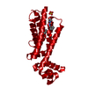
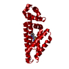
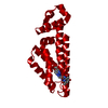
| Title | Structure of Transcriptional Regulatory Repressor Protein - EthR from Mycobacterium Tuberculosis in complex with N-((tetrahydrofuran-3-yl)methyl)pyrrolidine-1-carboxamide at 1.93A resolution | ||||||||||||
 Components Components | TetR-family transcriptional regulatory repressor protein | ||||||||||||
 Keywords Keywords | TRANSCRIPTION / EthR / represor / boosting effect | ||||||||||||
| Function / homology |  Function and homology information Function and homology informationtranscription cis-regulatory region binding / DNA-binding transcription factor activity / negative regulation of DNA-templated transcription Similarity search - Function | ||||||||||||
| Biological species |   Mycobacterium tuberculosis (bacteria) Mycobacterium tuberculosis (bacteria) | ||||||||||||
| Method |  X-RAY DIFFRACTION / SYNCHROTRON / MOLECULAR REPLACEMENT / molecular replacement / Resolution: 1.93 Å X-RAY DIFFRACTION / SYNCHROTRON / MOLECULAR REPLACEMENT / molecular replacement / Resolution: 1.93 Å | ||||||||||||
 Authors Authors | Blaszczyk, M. / Surade, S. / Nikiforov, P.O. / Abell, C. / Blundell, T.L. | ||||||||||||
| Funding support |  United Kingdom, United Kingdom,  United States, 3items United States, 3items
| ||||||||||||
 Citation Citation | Journal: ACS Chem. Biol. / Year: 2017 Title: Fragment-Sized EthR Inhibitors Exhibit Exceptionally Strong Ethionamide Boosting Effect in Whole-Cell Mycobacterium tuberculosis Assays. Authors: Nikiforov, P.O. / Blaszczyk, M. / Surade, S. / Boshoff, H.I. / Sajid, A. / Delorme, V. / Deboosere, N. / Brodin, P. / Baulard, A.R. / Barry, C.E. / Blundell, T.L. / Abell, C. | ||||||||||||
| History |
|
- Structure visualization
| Structure viewer | Molecule: MolmilJmol/JSmol |
|---|
- Downloads & links
-Download
| PDBx/mmCIF format | 5ip6.cif.gz | 51.5 KB | Display | PDBx/mmCIF format |
|---|---|---|---|---|
| PDB format | pdb5ip6.ent.gz | 36.2 KB | Display | PDB format |
| PDBx/mmJSON format | 5ip6.json.gz | Tree view | PDBx/mmJSON format | |
| Others |  Other downloads Other downloads |
-Validation report
| Arichive directory | https://data.pdbj.org/pub/pdb/validation_reports/ip/5ip6ftp://data.pdbj.org/pub/pdb/validation_reports/ip/5ip6 | HTTPS FTP |
|---|
-Related structure data
| Related structure data |  5ioyC  5iozC  5ipaC  5j1rC  5j1uC  5j1yC  5j3lC 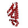 1t56S S: Starting model for refinement C: citing same article ( |
|---|---|
| Similar structure data |


-Links
 PDBj
PDBj- Assembly
| Deposited unit | 
| ||||||||
|---|---|---|---|---|---|---|---|---|---|
| 1 | 
| ||||||||
| Unit cell |
| ||||||||
| Components on special symmetry positions |
|
-Components
| #1: Protein | Mass: 23781.705 Da / Num. of mol.: 1 Source method: isolated from a genetically manipulated source Source: (gene. exp.) Mycobacterium tuberculosis (strain ATCC 25177 / H37Ra) (bacteria)Gene: ethR, MRA_3895 / Production host: |
|---|---|
| #2: Chemical | ChemComp-6C9 /   Mass: 198.262 Da / Num. of mol.: 1 / Source method: obtained synthetically / Formula: C10H18N2O2 Mass: 198.262 Da / Num. of mol.: 1 / Source method: obtained synthetically / Formula: C10H18N2O2 |
| #3: Water | ChemComp-HOH /  Mass: 18.015 Da / Num. of mol.: 32 / Source method: isolated from a natural source / Formula: H2O Mass: 18.015 Da / Num. of mol.: 32 / Source method: isolated from a natural source / Formula: H2O |
-Experimental details
-Experiment
| Experiment | Method: X-RAY DIFFRACTION / Number of used crystals: 1 |
|---|
- Sample preparation
| Crystal | Density Matthews: 2.59 Å3/Da / Density % sol: 52.47 % |
|---|---|
| Crystal grow | Temperature: 298 K / Method: vapor diffusion, sitting drop / pH: 6.5 / Details: Ammonium sulphate, Glycerol, MES / PH range: 6.3 - 6.5 |
-Data collection
| Diffraction | Mean temperature: 100 K | |||||||||||||||
|---|---|---|---|---|---|---|---|---|---|---|---|---|---|---|---|---|
| Diffraction source | Source: SYNCHROTRON / Site: Diamond / Beamline: I03 / Wavelength: 0.983 Å | |||||||||||||||
| Detector | Type: ADSC QUANTUM 315 / Detector: CCD / Date: Feb 9, 2013 | |||||||||||||||
| Radiation | Protocol: SINGLE WAVELENGTH / Monochromatic (M) / Laue (L): M / Scattering type: x-ray | |||||||||||||||
| Radiation wavelength | Wavelength: 0.983 Å / Relative weight: 1 | |||||||||||||||
| Reflection | Resolution: 1.91→85.73 Å / Num. obs: 39652 / % possible obs: 99.7 % / Redundancy: 6.4 % / CC1/2: 0.998 / Rmerge(I) obs: 0.1 / Rpim(I) all: 0.043 / Rrim(I) all: 0.11 / Net I/σ(I): 14.7 / Num. measured all: 252285 | |||||||||||||||
| Reflection shell |
|
-Phasing
| Phasing | Method: molecular replacement |
|---|
- Processing
| Software |
| |||||||||||||||||||||||||||||||||||||||||||||
|---|---|---|---|---|---|---|---|---|---|---|---|---|---|---|---|---|---|---|---|---|---|---|---|---|---|---|---|---|---|---|---|---|---|---|---|---|---|---|---|---|---|---|---|---|---|---|
| Refinement | Method to determine structure: MOLECULAR REPLACEMENT Starting model: 1T56 Resolution: 1.93→29.45 Å / Cor.coef. Fo:Fc: 0.942 / Cor.coef. Fo:Fc free: 0.934 / SU B: 3.143 / SU ML: 0.092 / SU R Cruickshank DPI: 0.1485 / Cross valid method: THROUGHOUT / σ(F): 0 / ESU R: 0.148 / ESU R Free: 0.136 Details: HYDROGENS HAVE BEEN USED IF PRESENT IN THE INPUT U VALUES : REFINED INDIVIDUALLY
| |||||||||||||||||||||||||||||||||||||||||||||
| Solvent computation | Ion probe radii: 0.8 Å / Shrinkage radii: 0.8 Å / VDW probe radii: 1.2 Å | |||||||||||||||||||||||||||||||||||||||||||||
| Displacement parameters | Biso max: 92.34 Å2 / Biso mean: 28.807 Å2 / Biso min: 9.95 Å2
| |||||||||||||||||||||||||||||||||||||||||||||
| Refinement step | Cycle: final / Resolution: 1.93→29.45 Å
| |||||||||||||||||||||||||||||||||||||||||||||
| Refine LS restraints |
| |||||||||||||||||||||||||||||||||||||||||||||
| LS refinement shell | Resolution: 1.928→1.978 Å / Total num. of bins used: 20
|
