[English] 日本語
Yorodumi
- PDB-5ngi: Structure of XcpQN012 -

+
Open data


ID or keywords:

Loading...

-
Basic information

Entry
Database: PDB / ID: 5ngi
TitleStructure of XcpQN012
ComponentsType II secretion system protein DType II secretion system
Keywords
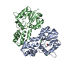
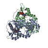
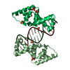
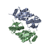
TRANSPORT PROTEIN / Secretion system II / Pseudomonas aeruginosa / secretin N domain
Function / homology
Function and homology information


protein secretion by the type II secretion system / type II protein secretion system complex / protein secretion / cell outer membrane / identical protein binding
Similarity search - Function
: / GspD-like, N0 domain / Ribosomal Protein S8; Chain: A, domain 1 - #120 / Type II secretion system protein GspD / GspD/PilQ family / Bacterial type II secretion system protein D signature. / Type II secretion system protein GspD, conserved site / NolW-like / Bacterial type II/III secretion system short domain / NolW-like superfamily ...: / GspD-like, N0 domain / Ribosomal Protein S8; Chain: A, domain 1 - #120 / Type II secretion system protein GspD / GspD/PilQ family / Bacterial type II secretion system protein D signature. / Type II secretion system protein GspD, conserved site / NolW-like / Bacterial type II/III secretion system short domain / NolW-like superfamily / Type II/III secretion system / Bacterial type II and III secretion system protein / Ribosomal Protein S8; Chain: A, domain 1 / 2-Layer Sandwich / Alpha Beta
Similarity search - Domain/homology
Type II secretion system protein D / Secretin XcpQ
Similarity search - Component
Biological speciesPseudomonas aeruginosa (bacteria)
MethodX-RAY DIFFRACTION / SYNCHROTRON / MOLECULAR REPLACEMENT / Resolution: 2.98 Å
AuthorsCambillau, C. / Roussel, A. / Trinh, T.N.
Funding support France, 1items
OrganizationGrant numberCountry
bourses excellence EIFFEL840686C France
Citation
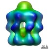
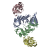
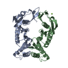
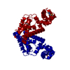
Journal: mBio / Year: 2017
Title: Unraveling the Self-Assembly of the XcpQ Secretin Periplasmic Domain Provides New Molecular Insights into Type II Secretion System Secreton Architecture and Dynamics.
Authors: Badreddine Douzi / Nhung T T Trinh / Sandra Michel-Souzy / Aline Desmyter / Geneviève Ball / Pascale Barbier / Artemis Kosta / Eric Durand / Katrina T Forest / Christian Cambillau / Alain ...Authors: Badreddine Douzi / Nhung T T Trinh / Sandra Michel-Souzy / Aline Desmyter / Geneviève Ball / Pascale Barbier / Artemis Kosta / Eric Durand / Katrina T Forest / Christian Cambillau / Alain Roussel / Romé Voulhoux /
Abstract: The type II secretion system (T2SS) releases large folded exoproteins across the envelope of many Gram-negative pathogens. This secretion process therefore requires specific gating, interacting, and ...The type II secretion system (T2SS) releases large folded exoproteins across the envelope of many Gram-negative pathogens. This secretion process therefore requires specific gating, interacting, and dynamics properties mainly operated by a bipartite outer membrane channel called secretin. We have a good understanding of the structure-function relationship of the pore-forming C-terminal domain of secretins. In contrast, the high flexibility of their periplasmic N-terminal domain has been an obstacle in obtaining the detailed structural information required to uncover its molecular function. In , the Xcp T2SS plays an important role in bacterial virulence by its capacity to deliver a large panel of toxins and degradative enzymes into the surrounding environment. Here, we revealed that the N-terminal domain of XcpQ secretin spontaneously self-assembled into a hexamer of dimers independently of its C-terminal domain. Furthermore, and by using multidisciplinary approaches, we elucidate the structural organization of the XcpQ N domain and demonstrate that secretin flexibility at interdimer interfaces is mandatory for its function. Bacterial secretins are large homooligomeric proteins constituting the outer membrane pore-forming element of several envelope-embedded nanomachines essential in bacterial survival and pathogenicity. They comprise a well-defined membrane-embedded C-terminal domain and a modular periplasmic N-terminal domain involved in substrate recruitment and connection with inner membrane components. We are studying the XcpQ secretin of the T2SS present in the pathogenic bacterium Our data highlight the ability of the XcpQ N-terminal domain to spontaneously oligomerize into a hexamer of dimers. Further experiments revealed that this domain adopts different conformations essential for the T2SS secretion process. These findings provide new insights into the functional understanding of bacterial T2SS secretins.
History
DepositionMar 17, 2017Deposition site: PDBE / Processing site: PDBE
Revision 1.0Jan 24, 2018Provider: repository / Type: Initial release
Revision 1.1Jan 31, 2018Group: Structure summary / Category: audit_author / struct / Item: _audit_author.name / _struct.title
Revision 1.2Jan 17, 2024Group: Data collection / Database references / Refinement description
Category: chem_comp_atom / chem_comp_bond ...chem_comp_atom / chem_comp_bond / database_2 / pdbx_initial_refinement_model
Item: _database_2.pdbx_DOI / _database_2.pdbx_database_accession

-
Structure visualization

Structure viewerMolecule:
MolmilJmol/JSmol

Downloads & links

-
Assembly

Deposited unit
A: Type II secretion system protein D
B: Type II secretion system protein D


Theoretical massNumber of molelcules
Total (without water)51,8702
Polymers51,8702
Non-polymers00
Water23413
1


  • Idetical with deposited unit
  • defined by author&software
TypeNameSymmetry operationNumber
identity operation1_555x,y,z1
Buried area1970 Å2
ΔGint-2 kcal/mol
Surface area20270 Å2
MethodPISA
Unit cell
Length a, b, c (Å)40.400, 122.250, 55.440
Angle α, β, γ (deg.)90.00, 109.06, 90.00
Int Tables number4
Space group name H-MP1211

-
Components

#1: Protein Type II secretion system protein D / Type II secretion system


Mass: 25935.201 Da / Num. of mol.: 2 / Fragment: N domain, UNP residues 79-319
Source method: isolated from a genetically manipulated source
Source: (gene. exp.) Pseudomonas aeruginosa (bacteria) / Gene: xcpQ1, PAMH19_1963 / Production host: Escherichia coli (E. coli) / References: UniProt: A0A0A8RG33, UniProt: P35818*PLUS
#2: Water ChemComp-HOH / water / Water


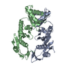
Mass: 18.015 Da / Num. of mol.: 13 / Source method: isolated from a natural source / Formula: H2O

-
Experimental details

-
Experiment

ExperimentMethod: X-RAY DIFFRACTION / Number of used crystals: 1

-
Sample preparation

CrystalDensity Matthews: 2.49 Å3/Da / Density % sol: 50.7 %
Crystal growTemperature: 273 K / Method: vapor diffusion, sitting drop / pH: 8.5
Details: 0.8M lithium chloride, 0.1M Tris-HCl, pH 8.5, 0.1M sodium acetate, and 32% PEG 4000 (w/v).

-
Data collection

DiffractionMean temperature: 100 K
Diffraction sourceSource: SYNCHROTRON / Site: SOLEIL / Beamline: PROXIMA 1 / Wavelength: 0.93 Å
DetectorType: DECTRIS EIGER X 16M / Detector: PIXEL / Date: Jan 30, 2017
RadiationProtocol: SINGLE WAVELENGTH / Monochromatic (M) / Laue (L): M / Scattering type: x-ray
Radiation wavelengthWavelength: 0.93 Å / Relative weight: 1
ReflectionResolution: 2.98→48.2 Å / Num. obs: 10400 / % possible obs: 99.3 % / Redundancy: 4.5 % / Biso Wilson estimate: 124.95 Å2 / CC1/2: 0.998 / Rmerge(I) obs: 0.058 / Net I/σ(I): 14.9
Reflection shellResolution: 2.98→3.16 Å / Redundancy: 4.4 % / Rmerge(I) obs: 0.7 / Mean I/σ(I) obs: 2 / Num. unique obs: 1659 / CC1/2: 0.982 / % possible all: 98.2

-
Processing

Software
NameVersionClassification
BUSTER2.10.1refinement
XDSdata reduction
XSCALEdata scaling
MOLREPphasing
RefinementMethod to determine structure: MOLECULAR REPLACEMENT
Starting model: 4E9J
Resolution: 2.98→48.16 Å / Cor.coef. Fo:Fc: 0.9539 / Cor.coef. Fo:Fc free: 0.9497 / Cross valid method: THROUGHOUT / σ(F): 0 / SU Rfree Blow DPI: 0.351
RfactorNum. reflection% reflectionSelection details
Rfree0.228 520 5 %RANDOM
Rwork0.2013 ---
obs0.2026 10394 99.63 %-
Displacement parametersBiso mean: 137.35 Å2
Baniso -1Baniso -2Baniso -3
1-15.8529 Å20 Å2-18.9322 Å2
2---2.1276 Å20 Å2
3----13.7253 Å2
Refine analyzeLuzzati coordinate error obs: 0.993 Å
Refinement stepCycle: 1 / Resolution: 2.98→48.16 Å
ProteinNucleic acidLigandSolventTotal
Num. atoms3022 0 0 13 3035
Refine LS restraints
Refine-IDTypeDev idealNumberRestraint functionWeight
X-RAY DIFFRACTIONt_bond_d0.013067HARMONIC2
X-RAY DIFFRACTIONt_angle_deg1.234211HARMONIC2
X-RAY DIFFRACTIONt_dihedral_angle_d970SINUSOIDAL2
X-RAY DIFFRACTIONt_incorr_chiral_ct
X-RAY DIFFRACTIONt_pseud_angle
X-RAY DIFFRACTIONt_trig_c_planes71HARMONIC2
X-RAY DIFFRACTIONt_gen_planes459HARMONIC5
X-RAY DIFFRACTIONt_it3067HARMONIC20
X-RAY DIFFRACTIONt_nbd
X-RAY DIFFRACTIONt_omega_torsion2.48
X-RAY DIFFRACTIONt_other_torsion23.42
X-RAY DIFFRACTIONt_improper_torsion
X-RAY DIFFRACTIONt_chiral_improper_torsion454SEMIHARMONIC5
X-RAY DIFFRACTIONt_sum_occupancies
X-RAY DIFFRACTIONt_utility_distance
X-RAY DIFFRACTIONt_utility_angle
X-RAY DIFFRACTIONt_utility_torsion
X-RAY DIFFRACTIONt_ideal_dist_contact3291SEMIHARMONIC4
LS refinement shellResolution: 2.98→3.33 Å / Total num. of bins used: 5
RfactorNum. reflection% reflection
Rfree0.3155 147 5 %
Rwork0.2464 2791 -
all0.2496 2938 -
obs--99.63 %
Refinement TLS params.

Method: refined / Refine-ID: X-RAY DIFFRACTION

IDL112)L122)L132)L222)L232)L332)S11 (Å °)S12 (Å °)S13 (Å °)S21 (Å °)S22 (Å °)S23 (Å °)S31 (Å °)S32 (Å °)S33 (Å °)T112)T122)T132)T222)T232)T332)Origin x (Å)Origin y (Å)Origin z (Å)
10.7860.0466-0.24376.61113.04994.2025-0.24560.43830.4411-0.1457-0.052-0.14970.1645-0.4190.2977-0.1018-0.06210.1183-0.0816-0.09450.0892-1.85571.140410.4417
25.74563.5132-1.45665.3785-1.25621.9366-0.2433-0.14640.25420.31120.0151-0.46490.43930.0270.2281-0.0389-0.04310.125-0.224-0.08720.07285.2789-5.354329.6399
Refinement TLS group
IDRefine-IDRefine TLS-IDSelection details
1X-RAY DIFFRACTION1{ A|* }
2X-RAY DIFFRACTION2{ B|* }

+
About Yorodumi

-
News

-
Feb 9, 2022. New format data for meta-information of EMDB entries

New format data for meta-information of EMDB entries

  • Version 3 of the EMDB header file is now the official format.
  • The previous official version 1.9 will be removed from the archive.

Related info.:EMDB header

External links:wwPDB to switch to version 3 of the EMDB data model

-
Aug 12, 2020. Covid-19 info

Covid-19 info

URL: https://pdbj.org/emnavi/covid19.php

New page: Covid-19 featured information page in EM Navigator.

Related info.:Covid-19 info / Mar 5, 2020. Novel coronavirus structure data

+
Mar 5, 2020. Novel coronavirus structure data

Novel coronavirus structure data

Related info.:Yorodumi Speices / Aug 12, 2020. Covid-19 info

External links:COVID-19 featured content - PDBj / Molecule of the Month (242):Coronavirus Proteases

+
Jan 31, 2019. EMDB accession codes are about to change! (news from PDBe EMDB page)

EMDB accession codes are about to change! (news from PDBe EMDB page)

  • The allocation of 4 digits for EMDB accession codes will soon come to an end. Whilst these codes will remain in use, new EMDB accession codes will include an additional digit and will expand incrementally as the available range of codes is exhausted. The current 4-digit format prefixed with “EMD-” (i.e. EMD-XXXX) will advance to a 5-digit format (i.e. EMD-XXXXX), and so on. It is currently estimated that the 4-digit codes will be depleted around Spring 2019, at which point the 5-digit format will come into force.
  • The EM Navigator/Yorodumi systems omit the EMD- prefix.

Related info.:Q: What is EMD? / ID/Accession-code notation in Yorodumi/EM Navigator

External links:EMDB Accession Codes are Changing Soon! / Contact to PDBj

+
Jul 12, 2017. Major update of PDB

Major update of PDB

  • wwPDB released updated PDB data conforming to the new PDBx/mmCIF dictionary.
  • This is a major update changing the version number from 4 to 5, and with Remediation, in which all the entries are updated.
  • In this update, many items about electron microscopy experimental information are reorganized (e.g. em_software).
  • Now, EM Navigator and Yorodumi are based on the updated data.

External links:wwPDB Remediation / Enriched Model Files Conforming to OneDep Data Standards Now Available in the PDB FTP Archive

-
Yorodumi

Thousand views of thousand structures

  • Yorodumi is a browser for structure data from EMDB, PDB, SASBDB, etc.
  • This page is also the successor to EM Navigator detail page, and also detail information page/front-end page for Omokage search.
  • The word "yorodu" (or yorozu) is an old Japanese word meaning "ten thousand". "mi" (miru) is to see.

Related info.:EMDB / PDB / SASBDB / Comparison of 3 databanks / Yorodumi Search / Aug 31, 2016. New EM Navigator & Yorodumi / Yorodumi Papers / Jmol/JSmol / Function and homology information / Changes in new EM Navigator and Yorodumi

Read more