 Movie
Movie Controller


+ Open data

- Basic information
| Entry | Database: PDB / ID: 7ner | ||||||
|---|---|---|---|---|---|---|---|
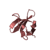
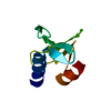
| Title | Crystal structure of the v-Src SH3 domain Q128R mutant | ||||||
 Components Components | v-Src SH3 domain | ||||||
 Keywords Keywords | PROTEIN BINDING / beta barrel / SH3 domain | ||||||
| Biological species |  | ||||||
| Method |  X-RAY DIFFRACTION / SYNCHROTRON / MOLECULAR REPLACEMENT / molecular replacement / Resolution: 1.55 Å X-RAY DIFFRACTION / SYNCHROTRON / MOLECULAR REPLACEMENT / molecular replacement / Resolution: 1.55 Å | ||||||
| Model details | Intertwined dimer | ||||||
 Authors Authors | Camara-Artigas, A. / Salinas-Garcia, M.C. | ||||||
| Funding support |  Spain, 1items Spain, 1items
| ||||||
 Citation Citation | Journal: Acta Crystallogr D Struct Biol / Year: 2021 Title: The impact of oncogenic mutations of the viral Src kinase on the structure and stability of the SH3 domain. Authors: Salinas-Garcia, M.C. / Plaza-Garrido, M. / Camara-Artigas, A. #1: Journal: Acta Crystallogr.,Sect.D / Year: 2021Title: The impact of oncogenic mutations of the viral Src kinase on the structure and stability of the SH3 domain Authors: Salinas-Garcia, M.C. / Plaza-Garrido, M. / Camara-Artigas, A. | ||||||
| History |
|
- Structure visualization
| Structure viewer | Molecule:  MolmilJmol/JSmol MolmilJmol/JSmol |
|---|
- Downloads & links
-Download
| PDBx/mmCIF format | 7ner.cif.gz | 50.9 KB | Display | PDBx/mmCIF format |
|---|---|---|---|---|
| PDB format | pdb7ner.ent.gz | 34.3 KB | Display | PDB format |
| PDBx/mmJSON format | 7ner.json.gz | Tree view | PDBx/mmJSON format | |
| Others |  Other downloads Other downloads |
-Validation report
| Summary document | 7ner_validation.pdf.gz | 422.4 KB | Display | wwPDB validaton report |
|---|---|---|---|---|
| Full document | 7ner_full_validation.pdf.gz | 422.4 KB | Display | |
| Data in XML | 7ner_validation.xml.gz | 4.8 KB | Display | |
| Data in CIF | 7ner_validation.cif.gz | 6 KB | Display | |
| Arichive directory | https://data.pdbj.org/pub/pdb/validation_reports/ne/7nerftp://data.pdbj.org/pub/pdb/validation_reports/ne/7ner | HTTPS FTP |
-Related structure data
| Related structure data | 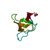 7nesC  7netC 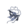 6xvnS S: Starting model for refinement C: citing same article ( |
|---|---|
| Similar structure data |


-Links
 PDBj
PDBj- Assembly
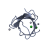
| Deposited unit | 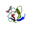
| ||||||||||||
|---|---|---|---|---|---|---|---|---|---|---|---|---|---|
| 1 |
| ||||||||||||
| Unit cell |
|
-Components
| #1: Protein | Mass: 6861.487 Da / Num. of mol.: 1 / Mutation: Q128R Source method: isolated from a genetically manipulated source Source: (gene. exp.)  |
|---|---|
| #2: Chemical | ChemComp-PG4 /   Mass: 194.226 Da / Num. of mol.: 1 / Source method: obtained synthetically / Formula: C8H18O5 / Comment: precipitant*YM Mass: 194.226 Da / Num. of mol.: 1 / Source method: obtained synthetically / Formula: C8H18O5 / Comment: precipitant*YM |
| #3: Chemical | ChemComp-SO4 /   Mass: 96.063 Da / Num. of mol.: 1 / Source method: obtained synthetically / Formula: SO4 Mass: 96.063 Da / Num. of mol.: 1 / Source method: obtained synthetically / Formula: SO4 |
| #4: Water | ChemComp-HOH /  Mass: 18.015 Da / Num. of mol.: 48 / Source method: isolated from a natural source / Formula: H2O Mass: 18.015 Da / Num. of mol.: 48 / Source method: isolated from a natural source / Formula: H2O |
| Has ligand of interest | N |
-Experimental details
-Experiment
| Experiment | Method: X-RAY DIFFRACTION / Number of used crystals: 1 |
|---|
- Sample preparation
| Crystal | Density Matthews: 1.79 Å3/Da / Density % sol: 31.37 % / Mosaicity: 0.09 ° |
|---|---|
| Crystal grow | Temperature: 283 K / Method: vapor diffusion, sitting drop / pH: 5.5 Details: 2.1 M ammonium sulfate, 5% PEG 200, 10% glycerol, 40 mM LiCl and 0.1M sodium acetate |
-Data collection
| Diffraction | Mean temperature: 100 K / Serial crystal experiment: N | |||||||||||||||||||||||||||
|---|---|---|---|---|---|---|---|---|---|---|---|---|---|---|---|---|---|---|---|---|---|---|---|---|---|---|---|---|
| Diffraction source | Source: SYNCHROTRON / Site: ESRF  / Beamline: ID30B / Wavelength: 0.9686 Å / Beamline: ID30B / Wavelength: 0.9686 Å | |||||||||||||||||||||||||||
| Detector | Type: DECTRIS PILATUS 6M / Detector: PIXEL / Date: Sep 30, 2020 | |||||||||||||||||||||||||||
| Radiation | Protocol: SINGLE WAVELENGTH / Monochromatic (M) / Laue (L): M / Scattering type: x-ray | |||||||||||||||||||||||||||
| Radiation wavelength | Wavelength: 0.9686 Å / Relative weight: 1 | |||||||||||||||||||||||||||
| Reflection | Resolution: 1.55→30.95 Å / Num. obs: 6888 / % possible obs: 98 % / Redundancy: 2.7 % / Biso Wilson estimate: 14.77 Å2 / CC1/2: 0.998 / Rmerge(I) obs: 0.054 / Rpim(I) all: 0.039 / Rrim(I) all: 0.067 / Net I/σ(I): 10.4 | |||||||||||||||||||||||||||
| Reflection shell | Diffraction-ID: 1 / Redundancy: 2.6 %
|
-Phasing
| Phasing | Method: molecular replacement |
|---|
- Processing
| Software |
| |||||||||||||||||||||||||||||||||||
|---|---|---|---|---|---|---|---|---|---|---|---|---|---|---|---|---|---|---|---|---|---|---|---|---|---|---|---|---|---|---|---|---|---|---|---|---|
| Refinement | Method to determine structure: MOLECULAR REPLACEMENT Starting model: 6XVN Resolution: 1.55→19.08 Å / SU ML: 0.0741 / Cross valid method: FREE R-VALUE / σ(F): 0.34 / Phase error: 18.649 Stereochemistry target values: GeoStd + Monomer Library + CDL v1.2
| |||||||||||||||||||||||||||||||||||
| Solvent computation | Shrinkage radii: 0.9 Å / VDW probe radii: 1.11 Å / Solvent model: FLAT BULK SOLVENT MODEL | |||||||||||||||||||||||||||||||||||
| Displacement parameters | Biso mean: 18.69 Å2 | |||||||||||||||||||||||||||||||||||
| Refinement step | Cycle: LAST / Resolution: 1.55→19.08 Å
| |||||||||||||||||||||||||||||||||||
| Refine LS restraints |
| |||||||||||||||||||||||||||||||||||
| LS refinement shell |
|
