 Movie
Movie Controller

[English] 日本語
 Yorodumi
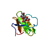
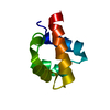
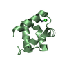
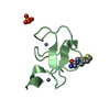
- PDB-1cka: STRUCTURAL BASIS FOR THE SPECIFIC INTERACTION OF LYSINE-CONTAININ... -
+ Open data

- Basic information
| Entry | Database: PDB / ID: 1cka | ||||||
|---|---|---|---|---|---|---|---|
| Title | STRUCTURAL BASIS FOR THE SPECIFIC INTERACTION OF LYSINE-CONTAINING PROLINE-RICH PEPTIDES WITH THE N-TERMINAL SH3 DOMAIN OF C-CRK | ||||||
 Components Components |
| ||||||
 Keywords Keywords | COMPLEX (ONCOGENE PROTEIN/PEPTIDE) / COMPLEX (ONCOGENE PROTEIN-PEPTIDE) / COMPLEX (ONCOGENE PROTEIN-PEPTIDE) complex | ||||||
| Function / homology |  Function and homology information Function and homology informationPTK6 Regulates RHO GTPases, RAS GTPase and MAP kinases / ARMS-mediated activation / MET activates RAP1 and RAC1 / response to hepatocyte growth factor / MET receptor recycling / helper T cell diapedesis / cerebellar neuron development / response to cholecystokinin / Downstream signal transduction / cellular response to endothelin ...PTK6 Regulates RHO GTPases, RAS GTPase and MAP kinases / ARMS-mediated activation / MET activates RAP1 and RAC1 / response to hepatocyte growth factor / MET receptor recycling / helper T cell diapedesis / cerebellar neuron development / response to cholecystokinin / Downstream signal transduction / cellular response to endothelin / postsynaptic specialization assembly / regulation of leukocyte migration / regulation of T cell migration / protein phosphorylated amino acid binding / p130Cas linkage to MAPK signaling for integrins / regulation of dendrite development / regulation of Rac protein signal transduction / negative regulation of wound healing / Regulation of signaling by CBL / regulation of intracellular signal transduction / response to peptide / Regulation of actin dynamics for phagocytic cup formation / response to yeast / reelin-mediated signaling pathway / positive regulation of skeletal muscle acetylcholine-gated channel clustering / VEGFA-VEGFR2 Pathway / negative regulation of cell motility / regulation of GTPase activity / negative regulation of natural killer cell mediated cytotoxicity / regulation of cell adhesion mediated by integrin / protein localization to membrane / positive regulation of smooth muscle cell migration / enzyme-linked receptor protein signaling pathway / cellular response to insulin-like growth factor stimulus / establishment of cell polarity / dendrite development / regulation of signal transduction / ephrin receptor signaling pathway / cellular response to transforming growth factor beta stimulus / positive regulation of Rac protein signal transduction / ephrin receptor binding / cytoskeletal protein binding / insulin-like growth factor receptor binding / phosphotyrosine residue binding / cellular response to nitric oxide / positive regulation of substrate adhesion-dependent cell spreading / signaling adaptor activity / SH2 domain binding / protein tyrosine kinase binding / regulation of actin cytoskeleton organization / cell chemotaxis / lipid metabolic process / hippocampus development / response to hydrogen peroxide / neuromuscular junction / cellular response to nerve growth factor stimulus / cerebral cortex development / SH3 domain binding / receptor tyrosine kinase binding / positive regulation of JNK cascade / neuron migration / kinase binding / regulation of cell shape / cell migration / actin cytoskeleton / signaling receptor complex adaptor activity / positive regulation of cell growth / actin cytoskeleton organization / scaffold protein binding / protein-macromolecule adaptor activity / cell population proliferation / protein domain specific binding / ubiquitin protein ligase binding / enzyme binding / protein-containing complex / membrane / plasma membrane / cytoplasm Similarity search - Function | ||||||
| Biological species |  | ||||||
| Method |  X-RAY DIFFRACTION / Resolution: 1.5 Å X-RAY DIFFRACTION / Resolution: 1.5 Å | ||||||
 Authors Authors | Wu, X. / Kuriyan, J. | ||||||
 Citation Citation | Journal: Structure / Year: 1995 Title: Structural basis for the specific interaction of lysine-containing proline-rich peptides with the N-terminal SH3 domain of c-Crk. Authors: Wu, X. / Knudsen, B. / Feller, S.M. / Zheng, J. / Sali, A. / Cowburn, D. / Hanafusa, H. / Kuriyan, J. | ||||||
| History |
|
- Structure visualization
| Structure viewer | Molecule: MolmilJmol/JSmol |
|---|
- Downloads & links
-Download
| PDBx/mmCIF format | 1cka.cif.gz | 34 KB | Display | PDBx/mmCIF format |
|---|---|---|---|---|
| PDB format | pdb1cka.ent.gz | 23.7 KB | Display | PDB format |
| PDBx/mmJSON format | 1cka.json.gz | Tree view | PDBx/mmJSON format | |
| Others |  Other downloads Other downloads |
-Validation report
| Arichive directory | https://data.pdbj.org/pub/pdb/validation_reports/ck/1ckaftp://data.pdbj.org/pub/pdb/validation_reports/ck/1cka | HTTPS FTP |
|---|
-Related structure data


-Links
 PDBj
PDBj


- Assembly
| Deposited unit | 
| ||||||||
|---|---|---|---|---|---|---|---|---|---|
| 1 |
| ||||||||
| Unit cell |
|
-Components
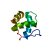
| #1: Protein | Mass: 6814.538 Da / Num. of mol.: 1 Source method: isolated from a genetically manipulated source Source: (gene. exp.)  |
|---|---|
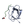
| #2: Protein/peptide | Mass: 1103.378 Da / Num. of mol.: 1 Source method: isolated from a genetically manipulated source |
| #3: Water | ChemComp-HOH /  Mass: 18.015 Da / Num. of mol.: 119 / Source method: isolated from a natural source / Formula: H2O Mass: 18.015 Da / Num. of mol.: 119 / Source method: isolated from a natural source / Formula: H2O |
-Experimental details
-Experiment
| Experiment | Method: X-RAY DIFFRACTION |
|---|
- Sample preparation
| Crystal | Density Matthews: 2.07 Å3/Da / Density % sol: 40.45 % | ||||||||||||||||||||||||||||||
|---|---|---|---|---|---|---|---|---|---|---|---|---|---|---|---|---|---|---|---|---|---|---|---|---|---|---|---|---|---|---|---|
| Crystal grow | *PLUS Temperature: 21 ℃ / pH: 4.6 / Method: vapor diffusion, hanging drop | ||||||||||||||||||||||||||||||
| Components of the solutions | *PLUS
|
-Data collection
| Diffraction source | Wavelength: 1.5418 |
|---|---|
| Detector | Type: RIGAKU RAXIS IIC / Detector: IMAGE PLATE / Date: Jul 16, 1994 |
| Radiation | Scattering type: x-ray |
| Radiation wavelength | Wavelength: 1.5418 Å / Relative weight: 1 |
| Reflection | Num. obs: 10512 / % possible obs: 81.3 % / Redundancy: 4.1 % |
| Reflection | *PLUS Highest resolution: 1.5 Å / Lowest resolution: 20 Å / Num. measured all: 66868 / Rmerge(I) obs: 0.04 |
| Reflection shell | *PLUS Highest resolution: 1.5 Å / Lowest resolution: 1.9 Å / % possible obs: 73.8 % / Mean I/σ(I) obs: 16.6 |
- Processing
| Software |
| ||||||||||||||||||||||||||||||||||||||||||||||||||||||||||||
|---|---|---|---|---|---|---|---|---|---|---|---|---|---|---|---|---|---|---|---|---|---|---|---|---|---|---|---|---|---|---|---|---|---|---|---|---|---|---|---|---|---|---|---|---|---|---|---|---|---|---|---|---|---|---|---|---|---|---|---|---|---|
| Refinement | Resolution: 1.5→6 Å / σ(F): 2 /
| ||||||||||||||||||||||||||||||||||||||||||||||||||||||||||||
| Displacement parameters | Biso mean: 8.3 Å2 | ||||||||||||||||||||||||||||||||||||||||||||||||||||||||||||
| Refinement step | Cycle: LAST / Resolution: 1.5→6 Å
| ||||||||||||||||||||||||||||||||||||||||||||||||||||||||||||
| Refine LS restraints |
| ||||||||||||||||||||||||||||||||||||||||||||||||||||||||||||
| Refinement | *PLUS | ||||||||||||||||||||||||||||||||||||||||||||||||||||||||||||
| Solvent computation | *PLUS | ||||||||||||||||||||||||||||||||||||||||||||||||||||||||||||
| Displacement parameters | *PLUS | ||||||||||||||||||||||||||||||||||||||||||||||||||||||||||||
| Refine LS restraints | *PLUS Type: x_angle_deg / Dev ideal: 1.9 |
