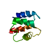
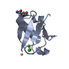
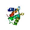
Mass: 9309.657 Da / Num. of mol.: 1 / Fragment: ste50 SAM domain (residues 32-107) Source method: isolated from a genetically manipulated source Source: (gene. exp.) Saccharomyces cerevisiae (brewer's yeast) Description: this protein fragment was cloned into the NdeI and BamHI sites of pET15b creating an aminoterminal hexahistidine tagged protein with an intervening thrombin protease cleavage site. After ...Description: this protein fragment was cloned into the NdeI and BamHI sites of pET15b creating an aminoterminal hexahistidine tagged protein with an intervening thrombin protease cleavage site. After thrombin treatment, the non-native residues GSH remained amino terminal to the native Ste50 sequence Gene: STE50 / Plasmid: pET15b / Species (production host): Escherichia coli / Production host: Escherichia coli BL21(DE3) (bacteria) / Strain (production host): BL21(DE3) / References: UniProt: P25344
-
Experimental details
-
Experiment
Experiment
Method: SOLUTION NMR
NMR experiment
Conditions-ID
Experiment-ID
Solution-ID
Type
1
1
1
3D 13C-separated NOESY
1
2
1
3D 15N-separated NOESY
NMR details
Text: This structure was determined using standard 3D heteronuclear techniques
-
Sample preparation
Details
Contents: 0.8 mM Ste50 SAM U-15N,13C, 20 mM sodium phosphate buffer, pH 7.8, 150 mM sodium chloride, 0.02 % sodium azide, 90% H2O, 10% D2O Solvent system: 90% H2O/10% D2O
Sample conditions
Ionic strength: 150 mM NaCl / pH: 7.8 / Pressure: 1 atm / Temperature: 293 K
-
NMR measurement
Radiation
Protocol: SINGLE WAVELENGTH / Monochromatic (M) / Laue (L): M
Radiation wavelength
Relative weight: 1
NMR spectrometer
Type
Manufacturer
Model
Field strength (MHz)
Spectrometer-ID
Bruker AVANCE
Bruker
AVANCE
600
1
Varian INOVA
Varian
INOVA
800
2
-
Processing
NMR software
Name
Version
Developer
Classification
NMRPipe
Delagio
processing
NMRView
5.2
Johnson
dataanalysis
CYANA
2
Guntert
structuresolution
XPLOR-NIH
2.9.9
refinement
Refinement
Method: simulated annealing / Software ordinal: 1 Details: Experimental observations: 615 intermolecular NOE distance restraints, 328 short range NOE distance restraints, 163 medium range NOE distance restraints, 149 long range NOE restraints, 42 ...Details: Experimental observations: 615 intermolecular NOE distance restraints, 328 short range NOE distance restraints, 163 medium range NOE distance restraints, 149 long range NOE restraints, 42 pairs of hydrogen bond distance restraints and 69 pairs of phi/psi dihedral angle restraints. An initial ensemble of 500 structures were calculated with CYANA 2.0. The top 25 structures with minimum restraint violations were refined in water using XPLOR-NIH and a protocol by C. Spronk. All 25 water refined structures had no NOE violations > 0.5 A and no dihedral violations > 5 degrees. For residues 35-100, the backbone RMSD of the ensemble is 0.75 +/- 0.14 A. Residues 28-32 and 101-107 are disordered.
NMR representative
Selection criteria: lowest energy
NMR ensemble
Conformers submitted total number: 1
+
About Yorodumi
-
News
-
Feb 9, 2022. New format data for meta-information of EMDB entries
New format data for meta-information of EMDB entries
Version 3 of the EMDB header file is now the official format.
The previous official version 1.9 will be removed from the archive.
In the structure databanks used in Yorodumi, some data are registered as the other names, "COVID-19 virus" and "2019-nCoV". Here are the details of the virus and the list of structure data.
Jan 31, 2019. EMDB accession codes are about to change! (news from PDBe EMDB page)
EMDB accession codes are about to change! (news from PDBe EMDB page)
The allocation of 4 digits for EMDB accession codes will soon come to an end. Whilst these codes will remain in use, new EMDB accession codes will include an additional digit and will expand incrementally as the available range of codes is exhausted. The current 4-digit format prefixed with “EMD-” (i.e. EMD-XXXX) will advance to a 5-digit format (i.e. EMD-XXXXX), and so on. It is currently estimated that the 4-digit codes will be depleted around Spring 2019, at which point the 5-digit format will come into force.
The EM Navigator/Yorodumi systems omit the EMD- prefix.
Related info.:Q: What is EMD? / ID/Accession-code notation in Yorodumi/EM Navigator
Yorodumi is a browser for structure data from EMDB, PDB, SASBDB, etc.
This page is also the successor to EM Navigator detail page, and also detail information page/front-end page for Omokage search.
The word "yorodu" (or yorozu) is an old Japanese word meaning "ten thousand". "mi" (miru) is to see.
Related info.:EMDB / PDB / SASBDB / Comparison of 3 databanks / Yorodumi Search / Aug 31, 2016. New EM Navigator & Yorodumi / Yorodumi Papers / Jmol/JSmol / Function and homology information / Changes in new EM Navigator and Yorodumi
 Movie
Movie Controller


 Open data
 Basic information
 Components
 Keywords
 Function and homology information
 Authors
 Citation
 Structure visualization
 Downloads & links
 Other downloads
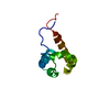


 PDBj
PDBj Assembly
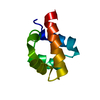

 Sample preparation
 Processing
 NMRPipe