Mass: 7186.223 Da / Num. of mol.: 1 / Fragment: SRC HOMOLOGY 3 DOMAIN Mutation: CIRCULAR PERMUTANT, CUT AT S19-P20, INS(MG-P20), INS(SG-T4) Source method: isolated from a genetically manipulated source Details: THIS IS A CIRCULAR PERMUTANT OF THE WT ALPHA-SPECTRIN SH3 SEQUENCE (PDB ENTRY WT-3D STRUCTURE: 1SGB). THE RESIDUE NUMBERS ARE AS IN THE WT SPECTRIN-SH3 DOMAIN (1SGB). THR 4 (N-TERMINUS) AND ...Details: THIS IS A CIRCULAR PERMUTANT OF THE WT ALPHA-SPECTRIN SH3 SEQUENCE (PDB ENTRY WT-3D STRUCTURE: 1SGB). THE RESIDUE NUMBERS ARE AS IN THE WT SPECTRIN-SH3 DOMAIN (1SGB). THR 4 (N-TERMINUS) AND ASP 62 (C-TERMINUS) OF THE WT-SH3 SEQUENCE ARE LINKED BY TWO ADDITIONAL RESIDUES (SER 2, GLY 3). THE CHAIN IS CLEAVED BETWEEN SER 19 AND PRO 20. TWO RESIDUES ARE ADDED AT THE NEW N-TERMINUS (MET 100, GLY 101). Source: (gene. exp.) Gallus gallus (chicken) / Organ: BRAIN / Plasmid: PET3D / Production host: Escherichia coli (E. coli) / References: UniProt: P07751
Mass: 18.015 Da / Num. of mol.: 70 / Source method: isolated from a natural source / Formula: H2O
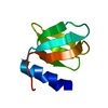
Sequence details
THIS IS A CIRCULAR PERMUTANT OF THE WT ALPHA-SPECTRIN SH3 SEQUENCE (PDB ENTRY WT-3D STRUCTURE: 1SGB) ...THIS IS A CIRCULAR PERMUTANT OF THE WT ALPHA-SPECTRIN SH3 SEQUENCE (PDB ENTRY WT-3D STRUCTURE: 1SGB). THE RESIDUE NUMBERS ARE AS IN THE WT SPECTRIN-SH3 DOMAIN (1SGB). THR 4 (N-TERMINUS) AND ASP 62 (C-TERMINUS) OF THE WT-SH3 SEQUENCE ARE LINKED BY TWO ADDITIONAL RESIDUES (SER 2, GLY 3). THE CHAIN IS CLEAVED BETWEEN SER 19 AND PRO 20. TWO RESIDUES ARE ADDED AT THE NEW N-TERMINUS (MET 100, GLY 101).
Resolution: 2.02→8 Å / σ(F): 1 Details: THE TWO C-TERMINAL RESIDUES (LYS 18, SER 19) ARE NOT VISIBLE IN THE ELECTRON DENSITY MAP. THERE IS NO SIDE CHAIN ELECTRON DENSITY FOR GLU 17.
Rfactor
Num. reflection
Rfree
0.29
-
Rwork
0.214
-
obs
0.214
5675
Displacement parameters
Biso mean: 29.88 Å2
Refinement step
Cycle: LAST / Resolution: 2.02→8 Å
Protein
Nucleic acid
Ligand
Solvent
Total
Num. atoms
484
0
0
70
554
Refine LS restraints
Refine-ID
Type
Dev ideal
X-RAY DIFFRACTION
x_bond_d
0.01
X-RAY DIFFRACTION
x_bond_d_na
X-RAY DIFFRACTION
x_bond_d_prot
X-RAY DIFFRACTION
x_angle_d
X-RAY DIFFRACTION
x_angle_d_na
X-RAY DIFFRACTION
x_angle_d_prot
X-RAY DIFFRACTION
x_angle_deg
1.577
X-RAY DIFFRACTION
x_angle_deg_na
X-RAY DIFFRACTION
x_angle_deg_prot
X-RAY DIFFRACTION
x_dihedral_angle_d
X-RAY DIFFRACTION
x_dihedral_angle_d_na
X-RAY DIFFRACTION
x_dihedral_angle_d_prot
X-RAY DIFFRACTION
x_improper_angle_d
1.262
X-RAY DIFFRACTION
x_improper_angle_d_na
X-RAY DIFFRACTION
x_improper_angle_d_prot
X-RAY DIFFRACTION
x_mcbond_it
X-RAY DIFFRACTION
x_mcangle_it
X-RAY DIFFRACTION
x_scbond_it
X-RAY DIFFRACTION
x_scangle_it
+
About Yorodumi
-
News
-
Feb 9, 2022. New format data for meta-information of EMDB entries
New format data for meta-information of EMDB entries
Version 3 of the EMDB header file is now the official format.
The previous official version 1.9 will be removed from the archive.
In the structure databanks used in Yorodumi, some data are registered as the other names, "COVID-19 virus" and "2019-nCoV". Here are the details of the virus and the list of structure data.
Jan 31, 2019. EMDB accession codes are about to change! (news from PDBe EMDB page)
EMDB accession codes are about to change! (news from PDBe EMDB page)
The allocation of 4 digits for EMDB accession codes will soon come to an end. Whilst these codes will remain in use, new EMDB accession codes will include an additional digit and will expand incrementally as the available range of codes is exhausted. The current 4-digit format prefixed with “EMD-” (i.e. EMD-XXXX) will advance to a 5-digit format (i.e. EMD-XXXXX), and so on. It is currently estimated that the 4-digit codes will be depleted around Spring 2019, at which point the 5-digit format will come into force.
The EM Navigator/Yorodumi systems omit the EMD- prefix.
Related info.:Q: What is EMD? / ID/Accession-code notation in Yorodumi/EM Navigator
Yorodumi is a browser for structure data from EMDB, PDB, SASBDB, etc.
This page is also the successor to EM Navigator detail page, and also detail information page/front-end page for Omokage search.
The word "yorodu" (or yorozu) is an old Japanese word meaning "ten thousand". "mi" (miru) is to see.
Related info.:EMDB / PDB / SASBDB / Comparison of 3 databanks / Yorodumi Search / Aug 31, 2016. New EM Navigator & Yorodumi / Yorodumi Papers / Jmol/JSmol / Function and homology information / Changes in new EM Navigator and Yorodumi
 Movie
Movie Controller

 Yorodumi
 Open data
 Basic information
 Components
 Keywords
 Function and homology information
 X-RAY DIFFRACTION / Resolution: 2.02 Å
  Authors
 Citation
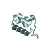
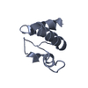
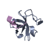
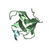
 Structure visualization
 Downloads & links
 Other downloads
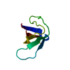


 PDBj
PDBj Assembly

 Mass: 18.015 Da / Num. of mol.: 70 / Source method: isolated from a natural source / Formula: H2O
 Sample preparation
 Processing