 Movie
Movie Controller

[English] 日本語
 Yorodumi
- PDB-4cc3: Complex of human Tuba C-terminal SH3 domain and Mena proline-rich... -
+ Open data

- Basic information
| Entry | Database: PDB / ID: 4cc3 | ||||||
|---|---|---|---|---|---|---|---|
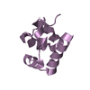
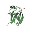
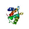
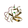
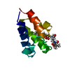
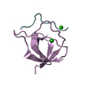
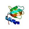
| Title | Complex of human Tuba C-terminal SH3 domain and Mena proline-rich peptide - H3 | ||||||
 Components Components |
| ||||||
 Keywords Keywords | STRUCTURAL PROTEIN / TUBA / SRC HOMOLOGY 3 / SH3 DOMAIN / MENA / NDPP-1 / PROLINE- RICH PEPTIDE / ACTIN CYTOSKELETON / CORTICAL TENSION | ||||||
| Function / homology |  Function and homology information Function and homology informationSignaling by ROBO receptors / postsynaptic cytoskeleton organization / actin polymerization-dependent cell motility / profilin binding / actin polymerization or depolymerization / Golgi stack / regulation of small GTPase mediated signal transduction / WW domain binding / CDC42 GTPase cycle / cilium assembly ...Signaling by ROBO receptors / postsynaptic cytoskeleton organization / actin polymerization-dependent cell motility / profilin binding / actin polymerization or depolymerization / Golgi stack / regulation of small GTPase mediated signal transduction / WW domain binding / CDC42 GTPase cycle / cilium assembly / cellular response to leukemia inhibitory factor / stress fiber / guanyl-nucleotide exchange factor activity / axon guidance / neural tube closure / actin filament organization / filopodium / SH3 domain binding / GABA-ergic synapse / cell-cell junction / regulation of cell shape / lamellipodium / actin cytoskeleton / presynapse / actin cytoskeleton organization / actin binding / cytoskeleton / postsynapse / nuclear body / intracellular signal transduction / focal adhesion / synapse / nucleolus / Golgi apparatus / nucleoplasm / plasma membrane / cytosol / cytoplasm Similarity search - Function | ||||||
| Biological species |  HOMO SAPIENS (human) HOMO SAPIENS (human) | ||||||
| Method |  X-RAY DIFFRACTION / SYNCHROTRON / MOLECULAR REPLACEMENT / Resolution: 1.97 Å X-RAY DIFFRACTION / SYNCHROTRON / MOLECULAR REPLACEMENT / Resolution: 1.97 Å | ||||||
 Authors Authors | Polle, L. / Rigano, L. / Julian, R. / Ireton, K. / Schubert, W.-D. | ||||||
 Citation Citation | Journal: Structure / Year: 2014 Title: Structural Details of Human Tuba Recruitment by Inlc of Listeria Monocytogenes Elucidate Bacterial Cell-Cell Spreading. Authors: Polle, L. / Rigano, L.A. / Julian, R. / Ireton, K. / Schubert, W. | ||||||
| History |
|
- Structure visualization
| Structure viewer | Molecule: MolmilJmol/JSmol |
|---|
- Downloads & links
-Download
| PDBx/mmCIF format | 4cc3.cif.gz | 137 KB | Display | PDBx/mmCIF format |
|---|---|---|---|---|
| PDB format | pdb4cc3.ent.gz | 109.3 KB | Display | PDB format |
| PDBx/mmJSON format | 4cc3.json.gz | Tree view | PDBx/mmJSON format | |
| Others |  Other downloads Other downloads |
-Validation report
| Arichive directory | https://data.pdbj.org/pub/pdb/validation_reports/cc/4cc3ftp://data.pdbj.org/pub/pdb/validation_reports/cc/4cc3 | HTTPS FTP |
|---|
-Related structure data
| Related structure data | 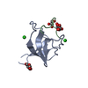 4cc2C  4cc4C  4cc7C  1zuuS C: citing same article ( S: Starting model for refinement |
|---|---|
| Similar structure data |


-Links
 PDBj
PDBj


- Assembly
| Deposited unit | 
| ||||||||
|---|---|---|---|---|---|---|---|---|---|
| 1 | 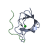
| ||||||||
| 2 | 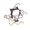
| ||||||||
| 3 | 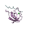
| ||||||||
| 4 | 
| ||||||||
| Unit cell |
|
-Components
| #1: Protein | Mass: 7712.617 Da / Num. of mol.: 4 / Fragment: C-TERMINAL SH3 DOMAIN, RESIDUES 1513-1577 Source method: isolated from a genetically manipulated source Source: (gene. exp.) HOMO SAPIENS (human) / Production host:  #2: Protein/peptide | Mass: 1163.321 Da / Num. of mol.: 4 / Fragment: PROLINE-RICH PEPTIDE, RESIDUES 547-558 / Source method: obtained synthetically / Source: (synth.) #3: Chemical |   Mass: 35.453 Da / Num. of mol.: 3 / Source method: obtained synthetically / Formula: Cl Mass: 35.453 Da / Num. of mol.: 3 / Source method: obtained synthetically / Formula: Cl#4: Chemical | ChemComp-PE4 / |   Mass: 354.436 Da / Num. of mol.: 1 / Source method: obtained synthetically / Formula: C16H34O8 / Comment: precipitant*YM Mass: 354.436 Da / Num. of mol.: 1 / Source method: obtained synthetically / Formula: C16H34O8 / Comment: precipitant*YM#5: Water | ChemComp-HOH / |  Mass: 18.015 Da / Num. of mol.: 148 / Source method: isolated from a natural source / Formula: H2O Mass: 18.015 Da / Num. of mol.: 148 / Source method: isolated from a natural source / Formula: H2OSequence details | THE FIRST TWO RESIDUES OF THE SEQUENCE GP ARE REMNANTS OF THE FUSION PROTEIN CLEAVAGE | |
|---|
-Experimental details
-Experiment
| Experiment | Method: X-RAY DIFFRACTION / Number of used crystals: 1 |
|---|
- Sample preparation
| Crystal | Density Matthews: 2.6 Å3/Da / Density % sol: 53 % / Description: NONE |
|---|---|
| Crystal grow | pH: 5.5 / Details: 2.5 M NACL, 0.1 M BIS-TRIS PH 5.5 |
-Data collection
| Diffraction | Mean temperature: 100 K |
|---|---|
| Diffraction source | Source: SYNCHROTRON / Site: SOLEIL  / Beamline: PROXIMA 1 / Wavelength: 0.98 / Beamline: PROXIMA 1 / Wavelength: 0.98 |
| Detector | Type: DECTRIS PILATUS 6M / Detector: PIXEL / Date: Dec 18, 2011 / Details: KIRKPATRICK-BAEZ PAIR OF BI-MORPH MIRRORS |
| Radiation | Protocol: SINGLE WAVELENGTH / Monochromatic (M) / Laue (L): M / Scattering type: x-ray |
| Radiation wavelength | Wavelength: 0.98 Å / Relative weight: 1 |
| Reflection | Resolution: 1.97→50 Å / Num. obs: 25648 / % possible obs: 99 % / Observed criterion σ(I): -3 / Redundancy: 4.2 % / Rmerge(I) obs: 0.04 / Net I/σ(I): 16.9 |
| Reflection shell | Resolution: 1.97→2.09 Å / Redundancy: 4.1 % / Rmerge(I) obs: 0.42 / Mean I/σ(I) obs: 3 / % possible all: 95.5 |
- Processing
| Software |
| ||||||||||||||||||||||||||||||||||||||||||||||||||||||||||||||||||||||||||||||||||||||||||||||||||||||||||||||||||||||||||||||||||||||||||||||||||||||||||||||||||||||||||||||||||||||
|---|---|---|---|---|---|---|---|---|---|---|---|---|---|---|---|---|---|---|---|---|---|---|---|---|---|---|---|---|---|---|---|---|---|---|---|---|---|---|---|---|---|---|---|---|---|---|---|---|---|---|---|---|---|---|---|---|---|---|---|---|---|---|---|---|---|---|---|---|---|---|---|---|---|---|---|---|---|---|---|---|---|---|---|---|---|---|---|---|---|---|---|---|---|---|---|---|---|---|---|---|---|---|---|---|---|---|---|---|---|---|---|---|---|---|---|---|---|---|---|---|---|---|---|---|---|---|---|---|---|---|---|---|---|---|---|---|---|---|---|---|---|---|---|---|---|---|---|---|---|---|---|---|---|---|---|---|---|---|---|---|---|---|---|---|---|---|---|---|---|---|---|---|---|---|---|---|---|---|---|---|---|---|---|
| Refinement | Method to determine structure: MOLECULAR REPLACEMENT Starting model: PDB ENTRY 1ZUU Resolution: 1.97→34.33 Å / Cor.coef. Fo:Fc: 0.968 / Cor.coef. Fo:Fc free: 0.957 / SU B: 5.721 / SU ML: 0.085 / Cross valid method: THROUGHOUT / ESU R: 0.033 / ESU R Free: 0.03 / Stereochemistry target values: MAXIMUM LIKELIHOOD Details: HYDROGENS HAVE BEEN ADDED IN THE RIDING POSITIONS. U VALUES WITH TLS ADDED
| ||||||||||||||||||||||||||||||||||||||||||||||||||||||||||||||||||||||||||||||||||||||||||||||||||||||||||||||||||||||||||||||||||||||||||||||||||||||||||||||||||||||||||||||||||||||
| Solvent computation | Ion probe radii: 0.8 Å / Shrinkage radii: 0.8 Å / VDW probe radii: 1.2 Å / Solvent model: BABINET MODEL WITH MASK | ||||||||||||||||||||||||||||||||||||||||||||||||||||||||||||||||||||||||||||||||||||||||||||||||||||||||||||||||||||||||||||||||||||||||||||||||||||||||||||||||||||||||||||||||||||||
| Displacement parameters | Biso mean: 47.781 Å2
| ||||||||||||||||||||||||||||||||||||||||||||||||||||||||||||||||||||||||||||||||||||||||||||||||||||||||||||||||||||||||||||||||||||||||||||||||||||||||||||||||||||||||||||||||||||||
| Refinement step | Cycle: LAST / Resolution: 1.97→34.33 Å
| ||||||||||||||||||||||||||||||||||||||||||||||||||||||||||||||||||||||||||||||||||||||||||||||||||||||||||||||||||||||||||||||||||||||||||||||||||||||||||||||||||||||||||||||||||||||
| Refine LS restraints |
|