 Movie
Movie Controller


+ Open data

- Basic information
| Entry | Database: PDB / ID: 3i2z | ||||||
|---|---|---|---|---|---|---|---|
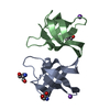
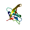
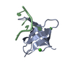
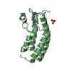
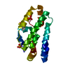
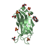
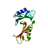
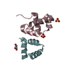
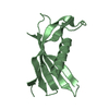
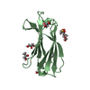
| Title | Structure of cold shock protein E from Salmonella typhimurium | ||||||
 Components Components | RNA chaperone, negative regulator of cspA transcription | ||||||
 Keywords Keywords | GENE REGULATION / BETA BARREL / DNA BINDING PROTEIN/TRANSCRIPTION / Cytoplasm | ||||||
| Function / homology |  Function and homology information Function and homology informationregulation of gene expression / nucleic acid binding / DNA binding / cytosol Similarity search - Function | ||||||
| Biological species |  Salmonella typhimurium (bacteria) Salmonella typhimurium (bacteria) | ||||||
| Method |  X-RAY DIFFRACTION / SYNCHROTRON / MOLECULAR REPLACEMENT / Resolution: 1.1 Å X-RAY DIFFRACTION / SYNCHROTRON / MOLECULAR REPLACEMENT / Resolution: 1.1 Å | ||||||
 Authors Authors | Morgan, H.P. / McNae, I. / Wear, M.A. / Gallagher, M. / Walkinshaw, M.D. | ||||||
 Citation Citation | Journal: Acta Crystallogr.,Sect.F / Year: 2009 Title: Crystallization and X-ray structure of cold-shock protein E from Salmonella typhimurium Authors: Morgan, H.P. / Wear, M.A. / McNae, I. / Gallagher, M.P. / Walkinshaw, M.D. | ||||||
| History |
|
- Structure visualization
| Structure viewer | Molecule: MolmilJmol/JSmol |
|---|
- Downloads & links
-Download
| PDBx/mmCIF format | 3i2z.cif.gz | 68.7 KB | Display | PDBx/mmCIF format |
|---|---|---|---|---|
| PDB format | pdb3i2z.ent.gz | 51.5 KB | Display | PDB format |
| PDBx/mmJSON format | 3i2z.json.gz | Tree view | PDBx/mmJSON format | |
| Others |  Other downloads Other downloads |
-Validation report
| Arichive directory | https://data.pdbj.org/pub/pdb/validation_reports/i2/3i2zftp://data.pdbj.org/pub/pdb/validation_reports/i2/3i2z | HTTPS FTP |
|---|
-Related structure data
| Related structure data | 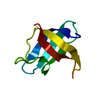 1mjcS S: Starting model for refinement |
|---|---|
| Similar structure data |


-Links
 PDBj
PDBj- Assembly
| Deposited unit | 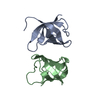
| ||||||||
|---|---|---|---|---|---|---|---|---|---|
| 1 |
| ||||||||
| 2 | 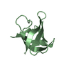
| ||||||||
| 3 | 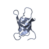
| ||||||||
| Unit cell |
|
-Components
| #1: Protein | Mass: 7685.595 Da / Num. of mol.: 2 Source method: isolated from a genetically manipulated source Source: (gene. exp.) Salmonella typhimurium (bacteria) / Strain: 1344 / Description: T7 expression - pET vector was used / Gene: cold shock protein E, cspE / Plasmid: pET28a_CspE / Production host: #2: Water | ChemComp-HOH / |  Mass: 18.015 Da / Num. of mol.: 100 / Source method: isolated from a natural source / Formula: H2O Mass: 18.015 Da / Num. of mol.: 100 / Source method: isolated from a natural source / Formula: H2O |
|---|
-Experimental details
-Experiment
| Experiment | Method: X-RAY DIFFRACTION / Number of used crystals: 1 |
|---|
- Sample preparation
| Crystal | Density Matthews: 2.03 Å3/Da / Density % sol: 39.4 % |
|---|---|
| Crystal grow | Temperature: 290 K / Method: vapor diffusion, hanging drop / pH: 9 Details: 28% PEG 20000, 0.05M AMPSO, 1% glycerol, pH 9, VAPOR DIFFUSION, HANGING DROP, temperature 290K |
-Data collection
| Diffraction | Mean temperature: 100 K |
|---|---|
| Diffraction source | Source: SYNCHROTRON / Site: SRS  / Beamline: PX10.1 / Wavelength: 1.074 Å / Beamline: PX10.1 / Wavelength: 1.074 Å |
| Detector | Type: MARMOSAIC 225 mm CCD / Detector: CCD / Date: Jun 28, 2006 / Details: Mirrors |
| Radiation | Monochromator: a double crystal Si(III,)with horizontal saggital focusing system Protocol: SINGLE WAVELENGTH / Monochromatic (M) / Laue (L): M / Scattering type: x-ray |
| Radiation wavelength | Wavelength: 1.074 Å / Relative weight: 1 |
| Reflection | Resolution: 1.1→45.36 Å / Num. obs: 47339 / % possible obs: 99.82 % / Observed criterion σ(F): 1.1 / Observed criterion σ(I): 1.1 / Redundancy: 5 % / Rmerge(I) obs: 0.047 / Num. measured all: 260904 |
| Reflection shell | Resolution: 1.1→1.16 Å / Redundancy: 3.8 % / Rmerge(I) obs: 0.392 / Mean I/σ(I) obs: 2.8 / Num. unique all: 7261 / Rsym value: 0.41 / % possible all: 100 |
- Processing
| Software |
| ||||||||||||||||||||||||||||||||||||||||||||||||||||||||||||||||||||||
|---|---|---|---|---|---|---|---|---|---|---|---|---|---|---|---|---|---|---|---|---|---|---|---|---|---|---|---|---|---|---|---|---|---|---|---|---|---|---|---|---|---|---|---|---|---|---|---|---|---|---|---|---|---|---|---|---|---|---|---|---|---|---|---|---|---|---|---|---|---|---|---|
| Refinement | Method to determine structure: MOLECULAR REPLACEMENT Starting model: PDB code 1MJC; A decapeptide (residues 12-21) from the E. coli cold shock protein A Resolution: 1.1→11.21 Å / Cor.coef. Fo:Fc: 0.961 / Cor.coef. Fo:Fc free: 0.946 / SU B: 1.514 / SU ML: 0.033 / Cross valid method: THROUGHOUT / ESU R: 0.037 / ESU R Free: 0.041 / Stereochemistry target values: MAXIMUM LIKELIHOOD
| ||||||||||||||||||||||||||||||||||||||||||||||||||||||||||||||||||||||
| Solvent computation | Ion probe radii: 0.8 Å / Shrinkage radii: 0.8 Å / VDW probe radii: 1.2 Å / Solvent model: MASK | ||||||||||||||||||||||||||||||||||||||||||||||||||||||||||||||||||||||
| Displacement parameters | Biso mean: 18.862 Å2
| ||||||||||||||||||||||||||||||||||||||||||||||||||||||||||||||||||||||
| Refinement step | Cycle: LAST / Resolution: 1.1→11.21 Å
| ||||||||||||||||||||||||||||||||||||||||||||||||||||||||||||||||||||||
| Refine LS restraints |
| ||||||||||||||||||||||||||||||||||||||||||||||||||||||||||||||||||||||
| LS refinement shell | Resolution: 1.1→1.129 Å / Total num. of bins used: 20
|
