 Movie
Movie Controller

[English] 日本語
 Yorodumi
- PDB-1fse: CRYSTAL STRUCTURE OF THE BACILLUS SUBTILIS REGULATORY PROTEIN GERE -
+ Open data

- Basic information
| Entry | Database: PDB / ID: 1fse | ||||||
|---|---|---|---|---|---|---|---|
| Title | CRYSTAL STRUCTURE OF THE BACILLUS SUBTILIS REGULATORY PROTEIN GERE | ||||||
 Components Components | GERE | ||||||
 Keywords Keywords | TRANSCRIPTION / Helix-turn-helix DNA-binding protein transcriptional regulator | ||||||
| Function / homology |  Function and homology information Function and homology informationregulation of DNA-templated transcription / DNA-templated transcription / DNA binding Similarity search - Function | ||||||
| Biological species |  | ||||||
| Method |  X-RAY DIFFRACTION / SYNCHROTRON / MAD / Resolution: 2.05 Å X-RAY DIFFRACTION / SYNCHROTRON / MAD / Resolution: 2.05 Å | ||||||
 Authors Authors | Ducros, V.M.-A. / Lewis, R.J. / Verma, C.S. / Dodson, E.J. / Leonard, G. / Turkenburg, J.P. / Murshudov, G.N. / Wilkinson, A.J. / Brannigan, J.A. | ||||||
 Citation Citation | Journal: J.Mol.Biol. / Year: 2001 Title: Crystal structure of GerE, the ultimate transcriptional regulator of spore formation in Bacillus subtilis. Authors: Ducros, V.M. / Lewis, R.J. / Verma, C.S. / Dodson, E.J. / Leonard, G. / Turkenburg, J.P. / Murshudov, G.N. / Wilkinson, A.J. / Brannigan, J.A. #1: Journal: Acta Crystallogr.,Sect.D / Year: 1998Title: Bacillus subtilis regulatory protein GerE Authors: Ducros, V. / Brannigan, J.A. / Lewis, R.J. / Wilkinson, A.J. | ||||||
| History |
|
- Structure visualization
| Structure viewer | Molecule: MolmilJmol/JSmol |
|---|
- Downloads & links
-Download
| PDBx/mmCIF format | 1fse.cif.gz | 93.5 KB | Display | PDBx/mmCIF format |
|---|---|---|---|---|
| PDB format | pdb1fse.ent.gz | 73.5 KB | Display | PDB format |
| PDBx/mmJSON format | 1fse.json.gz | Tree view | PDBx/mmJSON format | |
| Others |  Other downloads Other downloads |
-Validation report
| Arichive directory | https://data.pdbj.org/pub/pdb/validation_reports/fs/1fseftp://data.pdbj.org/pub/pdb/validation_reports/fs/1fse | HTTPS FTP |
|---|
-Related structure data
| Similar structure data |
|---|


-Links
 PDBj
PDBj- Assembly
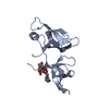
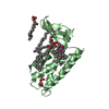
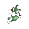
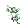
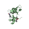
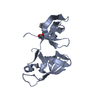
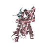
| Deposited unit | 
| ||||||||
|---|---|---|---|---|---|---|---|---|---|
| 1 | 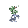
| ||||||||
| 2 | 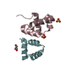
| ||||||||
| 3 | 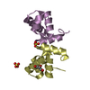
| ||||||||
| Unit cell |
| ||||||||
| Details | There six monomers in the asymmetric unit arranged as three pairs of dimers (A and B, C and F, D and E) |
-Components
| #1: Protein | Mass: 8603.060 Da / Num. of mol.: 6 Source method: isolated from a genetically manipulated source Source: (gene. exp.) #2: Chemical |   Mass: 92.094 Da / Num. of mol.: 3 / Source method: obtained synthetically / Formula: C3H8O3 Mass: 92.094 Da / Num. of mol.: 3 / Source method: obtained synthetically / Formula: C3H8O3#3: Chemical | ChemComp-SO4 /   Mass: 96.063 Da / Num. of mol.: 4 / Source method: obtained synthetically / Formula: SO4 Mass: 96.063 Da / Num. of mol.: 4 / Source method: obtained synthetically / Formula: SO4#4: Water | ChemComp-HOH / |  Mass: 18.015 Da / Num. of mol.: 322 / Source method: isolated from a natural source / Formula: H2O Mass: 18.015 Da / Num. of mol.: 322 / Source method: isolated from a natural source / Formula: H2O |
|---|
-Experimental details
-Experiment
| Experiment | Method: X-RAY DIFFRACTION / Number of used crystals: 1 |
|---|
- Sample preparation
| Crystal | Density Matthews: 2.3 Å3/Da / Density % sol: 46.06 % | ||||||||||||||||||||||||||||||
|---|---|---|---|---|---|---|---|---|---|---|---|---|---|---|---|---|---|---|---|---|---|---|---|---|---|---|---|---|---|---|---|
| Crystal grow | Method: vapor diffusion, hanging drop / pH: 5 Details: Peg4000, sodium acetate, lithium or ammonium sulfate, pH 5, VAPOR DIFFUSION, HANGING DROP | ||||||||||||||||||||||||||||||
| Crystal grow | *PLUS Temperature: 291 K | ||||||||||||||||||||||||||||||
| Components of the solutions | *PLUS
|
-Data collection
| Diffraction | Mean temperature: 100 K |
|---|---|
| Diffraction source | Source: SYNCHROTRON / Site: SRS  / Beamline: PX9.6 / Wavelength: 0.87 / Beamline: PX9.6 / Wavelength: 0.87 |
| Detector | Type: ADSC / Detector: IMAGE PLATE / Date: Feb 15, 1998 |
| Radiation | Protocol: SINGLE WAVELENGTH / Monochromatic (M) / Laue (L): M / Scattering type: x-ray |
| Radiation wavelength | Wavelength: 0.87 Å / Relative weight: 1 |
| Reflection | Resolution: 2.05→15 Å / Num. all: 25472 / Num. obs: 25472 / % possible obs: 99 % / Observed criterion σ(F): 0 / Observed criterion σ(I): 0 / Redundancy: 3.11 % / Biso Wilson estimate: 32 Å2 / Rmerge(I) obs: 0.068 / Net I/σ(I): 11.53 |
| Reflection shell | Resolution: 2.05→2.1 Å / Redundancy: 2.83 % / Rmerge(I) obs: 0.39 / Mean I/σ(I) obs: 4.07 / Num. unique all: 2421 / % possible all: 95.5 |
| Reflection | *PLUS Num. obs: 29311 / Num. measured all: 91541 |
| Reflection shell | *PLUS % possible obs: 95.5 % |
- Processing
| Software |
| ||||||||||||||||||||
|---|---|---|---|---|---|---|---|---|---|---|---|---|---|---|---|---|---|---|---|---|---|
| Refinement | Method to determine structure: MAD / Resolution: 2.05→20 Å / σ(F): 0 / σ(I): 0 / Stereochemistry target values: Engh & Huber Details: Used Translation, Libration and Screw motion (TLS) approach to refinement
| ||||||||||||||||||||
| Refinement step | Cycle: LAST / Resolution: 2.05→20 Å
| ||||||||||||||||||||
| Refine LS restraints |
|
