 Movie
Movie Controller

[English] 日本語
 Yorodumi
- PDB-1mjc: CRYSTAL STRUCTURE OF CSPA, THE MAJOR COLD SHOCK PROTEIN OF ESCHER... -
+ Open data

- Basic information
| Entry | Database: PDB / ID: 1mjc | ||||||
|---|---|---|---|---|---|---|---|
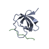
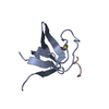
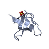
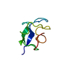
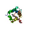
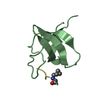
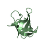
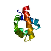
| Title | CRYSTAL STRUCTURE OF CSPA, THE MAJOR COLD SHOCK PROTEIN OF ESCHERICHIA COLI | ||||||
 Components Components | MAJOR COLD-SHOCK PROTEIN 7.4 | ||||||
 Keywords Keywords | TRANSCRIPTION REGULATION | ||||||
| Function / homology |  Function and homology information Function and homology informationnegative regulation of termination of DNA-templated transcription / transcription antitermination factor activity, RNA binding / response to cold / single-stranded DNA binding / regulation of gene expression / nucleic acid binding / single-stranded RNA binding / positive regulation of DNA-templated transcription / DNA binding / RNA binding / cytosol Similarity search - Function | ||||||
| Biological species |  | ||||||
| Method |  X-RAY DIFFRACTION / Resolution: 2 Å X-RAY DIFFRACTION / Resolution: 2 Å | ||||||
 Authors Authors | Schindelin, H. / Heinemann, U. | ||||||
 Citation Citation | Journal: Proc.Natl.Acad.Sci.USA / Year: 1994 Title: Crystal structure of CspA, the major cold shock protein of Escherichia coli. Authors: Schindelin, H. / Jiang, W. / Inouye, M. / Heinemann, U. #1: Journal: Nature / Year: 1993Title: Universal Nucleic Acid-Binding Domain Revealed by Crystal Structure of the B. Subtilis Major Cold Shock Protein Authors: Schindelin, H. / Marahiel, M. / Heinemann, U. #2: Journal: Proc.Natl.Acad.Sci.USA / Year: 1990Title: Major Cold Shock Protein of Escherichia Coli Authors: Goldstein, J. / Pollitt, N.S. / Inouye, M. | ||||||
| History |
|
- Structure visualization
| Structure viewer | Molecule: MolmilJmol/JSmol |
|---|
- Downloads & links
-Download
| PDBx/mmCIF format | 1mjc.cif.gz | 23.8 KB | Display | PDBx/mmCIF format |
|---|---|---|---|---|
| PDB format | pdb1mjc.ent.gz | 15.2 KB | Display | PDB format |
| PDBx/mmJSON format | 1mjc.json.gz | Tree view | PDBx/mmJSON format | |
| Others |  Other downloads Other downloads |
-Validation report
| Arichive directory | https://data.pdbj.org/pub/pdb/validation_reports/mj/1mjcftp://data.pdbj.org/pub/pdb/validation_reports/mj/1mjc | HTTPS FTP |
|---|
-Related structure data
| Similar structure data |
|---|


-Links
 PDBj
PDBj- Assembly
| Deposited unit | 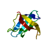
| ||||||||
|---|---|---|---|---|---|---|---|---|---|
| 1 |
| ||||||||
| Unit cell |
|
-Components
| #1: Protein | Mass: 7280.057 Da / Num. of mol.: 1 Source method: isolated from a genetically manipulated source Source: (gene. exp.) |
|---|---|
| #2: Water | ChemComp-HOH /  Mass: 18.015 Da / Num. of mol.: 36 / Source method: isolated from a natural source / Formula: H2O Mass: 18.015 Da / Num. of mol.: 36 / Source method: isolated from a natural source / Formula: H2O |
-Experimental details
-Experiment
| Experiment | Method: X-RAY DIFFRACTION |
|---|
- Sample preparation
| Crystal | Density Matthews: 2 Å3/Da / Density % sol: 38.4 % | ||||||||||||||||||||||||||||||
|---|---|---|---|---|---|---|---|---|---|---|---|---|---|---|---|---|---|---|---|---|---|---|---|---|---|---|---|---|---|---|---|
| Crystal grow | *PLUS pH: 7.5 / Method: vapor diffusion | ||||||||||||||||||||||||||||||
| Components of the solutions | *PLUS
|
-Data collection
| Radiation | Scattering type: x-ray |
|---|---|
| Radiation wavelength | Relative weight: 1 |
| Reflection | *PLUS Highest resolution: 2 Å / Num. obs: 3785 / % possible obs: 88.9 % / Num. measured all: 15912 / Rmerge(I) obs: 0.063 |
| Reflection shell | *PLUS Highest resolution: 2 Å / Lowest resolution: 2.13 Å / % possible obs: 87.8 % / Rmerge(I) obs: 0.113 / Mean I/σ(I) obs: 5.99 |
- Processing
| Software |
| ||||||||||||||||||||||||||||||||||||||||||||||||||||||||||||||||||||||||||||||||
|---|---|---|---|---|---|---|---|---|---|---|---|---|---|---|---|---|---|---|---|---|---|---|---|---|---|---|---|---|---|---|---|---|---|---|---|---|---|---|---|---|---|---|---|---|---|---|---|---|---|---|---|---|---|---|---|---|---|---|---|---|---|---|---|---|---|---|---|---|---|---|---|---|---|---|---|---|---|---|---|---|---|
| Refinement | Resolution: 2→10 Å / σ(F): 1 /
| ||||||||||||||||||||||||||||||||||||||||||||||||||||||||||||||||||||||||||||||||
| Refinement step | Cycle: LAST / Resolution: 2→10 Å
| ||||||||||||||||||||||||||||||||||||||||||||||||||||||||||||||||||||||||||||||||
| Refine LS restraints |
| ||||||||||||||||||||||||||||||||||||||||||||||||||||||||||||||||||||||||||||||||
| Refinement | *PLUS Rfactor obs: 0.187 / Rfactor Rwork: 0.187 | ||||||||||||||||||||||||||||||||||||||||||||||||||||||||||||||||||||||||||||||||
| Solvent computation | *PLUS | ||||||||||||||||||||||||||||||||||||||||||||||||||||||||||||||||||||||||||||||||
| Displacement parameters | *PLUS | ||||||||||||||||||||||||||||||||||||||||||||||||||||||||||||||||||||||||||||||||
| Refine LS restraints | *PLUS
|
