 Movie
Movie Controller

[English] 日本語
 Yorodumi
- PDB-6xvn: Crystal structure of c-Src SH3 domain without ATCUN motif: monomer 1 -
+ Open data

- Basic information
| Entry | Database: PDB / ID: 6xvn | ||||||
|---|---|---|---|---|---|---|---|
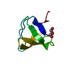
| Title | Crystal structure of c-Src SH3 domain without ATCUN motif: monomer 1 | ||||||
 Components Components | Proto-oncogene tyrosine-protein kinase Src | ||||||
 Keywords Keywords | PROTEIN BINDING / beta barrel / SH3 domain | ||||||
| Function / homology |  Function and homology information Function and homology informationSignaling by ERBB2 / Nuclear signaling by ERBB4 / Signaling by SCF-KIT / Regulation of KIT signaling / Signaling by EGFR / GAB1 signalosome / Regulation of gap junction activity / FCGR activation / PECAM1 interactions / Co-stimulation by CD28 ...Signaling by ERBB2 / Nuclear signaling by ERBB4 / Signaling by SCF-KIT / Regulation of KIT signaling / Signaling by EGFR / GAB1 signalosome / Regulation of gap junction activity / FCGR activation / PECAM1 interactions / Co-stimulation by CD28 / Co-inhibition by CTLA4 / EPHA-mediated growth cone collapse / Ephrin signaling / G alpha (i) signalling events / GP1b-IX-V activation signalling / Thrombin signalling through proteinase activated receptors (PARs) / VEGFR2 mediated cell proliferation / RET signaling / Receptor Mediated Mitophagy / ADP signalling through P2Y purinoceptor 1 / RAF activation / PIP3 activates AKT signaling / EPH-ephrin mediated repulsion of cells / PI5P, PP2A and IER3 Regulate PI3K/AKT Signaling / : / Downstream signal transduction / Downregulation of ERBB4 signaling / Cyclin D associated events in G1 / Regulation of RUNX3 expression and activity / Degradation of CDH1 / MAP2K and MAPK activation / Integrin signaling / GRB2:SOS provides linkage to MAPK signaling for Integrins / : / MET activates PTK2 signaling / Extra-nuclear estrogen signaling / EPHB-mediated forward signaling / p130Cas linkage to MAPK signaling for integrins / VEGFA-VEGFR2 Pathway / connexin binding / negative regulation of intrinsic apoptotic signaling pathway / progesterone receptor signaling pathway / immune system process / negative regulation of extrinsic apoptotic signaling pathway / non-specific protein-tyrosine kinase / non-membrane spanning protein tyrosine kinase activity / epidermal growth factor receptor signaling pathway / cell-cell junction / cell junction / protein tyrosine kinase activity / protein phosphatase binding / cytoskeleton / cell differentiation / cell adhesion / regulation of cell cycle / endosome membrane / mitochondrial inner membrane / signaling receptor binding / focal adhesion / heme binding / perinuclear region of cytoplasm / protein-containing complex / ATP binding / membrane / nucleus / plasma membrane / cytosol Similarity search - Function | ||||||
| Biological species |  | ||||||
| Method |  X-RAY DIFFRACTION / SYNCHROTRON / MOLECULAR REPLACEMENT / molecular replacement / Resolution: 1.7 Å X-RAY DIFFRACTION / SYNCHROTRON / MOLECULAR REPLACEMENT / molecular replacement / Resolution: 1.7 Å | ||||||
| Model details | dimer | ||||||
 Authors Authors | Camara-Artigas, A. / Plaza-Garrido, M. | ||||||
| Funding support |  Spain, 1items Spain, 1items
| ||||||
 Citation Citation | Journal: J.Biol.Inorg.Chem. / Year: 2020 Title: The effect of an engineered ATCUN motif on the structure and biophysical properties of the SH3 domain of c-Src tyrosine kinase. Authors: Plaza-Garrido, M. / Salinas-Garcia, M.C. / Martinez, J.C. / Camara-Artigas, A. | ||||||
| History |
|
- Structure visualization
| Structure viewer | Molecule: MolmilJmol/JSmol |
|---|
- Downloads & links
-Download
| PDBx/mmCIF format | 6xvn.cif.gz | 79.6 KB | Display | PDBx/mmCIF format |
|---|---|---|---|---|
| PDB format | pdb6xvn.ent.gz | 59.9 KB | Display | PDB format |
| PDBx/mmJSON format | 6xvn.json.gz | Tree view | PDBx/mmJSON format | |
| Others |  Other downloads Other downloads |
-Validation report
| Arichive directory | https://data.pdbj.org/pub/pdb/validation_reports/xv/6xvnftp://data.pdbj.org/pub/pdb/validation_reports/xv/6xvn | HTTPS FTP |
|---|
-Related structure data
| Related structure data | 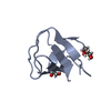 6xvmC 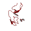 6xvoC 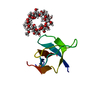 6xx2C 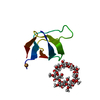 6xx3C 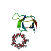 6xx4C 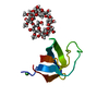 6xx5C  4jz4S S: Starting model for refinement C: citing same article ( |
|---|---|
| Similar structure data |


-Links
 PDBj
PDBj- Assembly
| Deposited unit | 
| ||||||||
|---|---|---|---|---|---|---|---|---|---|
| 1 | 
| ||||||||
| 2 | 
| ||||||||
| Unit cell |
|
-Components
| #1: Protein | Mass: 8852.580 Da / Num. of mol.: 2 Source method: isolated from a genetically manipulated source Source: (gene. exp.)  References: UniProt: P00523, non-specific protein-tyrosine kinase #2: Water | ChemComp-HOH / |  Mass: 18.015 Da / Num. of mol.: 125 / Source method: isolated from a natural source / Formula: H2O Mass: 18.015 Da / Num. of mol.: 125 / Source method: isolated from a natural source / Formula: H2O |
|---|
-Experimental details
-Experiment
| Experiment | Method: X-RAY DIFFRACTION / Number of used crystals: 1 |
|---|
- Sample preparation
| Crystal | Mosaicity: 0.13 ° |
|---|---|
| Crystal grow | Temperature: 298 K / Method: vapor diffusion, sitting drop / pH: 8 / Details: 2.5 ammonium sulfate, 0.1 TRIS |
-Data collection
| Diffraction | Mean temperature: 100 K / Serial crystal experiment: N | ||||||||||||||||||||||||||||||
|---|---|---|---|---|---|---|---|---|---|---|---|---|---|---|---|---|---|---|---|---|---|---|---|---|---|---|---|---|---|---|---|
| Diffraction source | Source: SYNCHROTRON / Site: ALBA / Beamline: XALOC / Wavelength: 0.97911 Å | ||||||||||||||||||||||||||||||
| Detector | Type: DECTRIS PILATUS 6M / Detector: PIXEL / Date: Mar 14, 2019 | ||||||||||||||||||||||||||||||
| Radiation | Protocol: SINGLE WAVELENGTH / Monochromatic (M) / Laue (L): M / Scattering type: x-ray | ||||||||||||||||||||||||||||||
| Radiation wavelength | Wavelength: 0.97911 Å / Relative weight: 1 | ||||||||||||||||||||||||||||||
| Reflection | Resolution: 1.7→19.13 Å / Num. obs: 15912 / % possible obs: 82.7 % / Redundancy: 2.7 % / CC1/2: 0.996 / Rmerge(I) obs: 0.052 / Rpim(I) all: 0.038 / Rrim(I) all: 0.065 / Net I/σ(I): 12.8 | ||||||||||||||||||||||||||||||
| Reflection shell | Diffraction-ID: 1
|
-Phasing
| Phasing | Method: molecular replacement |
|---|
- Processing
| Software |
| |||||||||||||||||||||||||||||||||||||||||||||||||||||||||||||||||||||||||||||||||||||||||||||||||||||||||||||||||||||||||||||||||||||||||||||||||||||||||||||||||||||||||||||||||||||||||||||||||||||||||||||||||||||||||||||||||
|---|---|---|---|---|---|---|---|---|---|---|---|---|---|---|---|---|---|---|---|---|---|---|---|---|---|---|---|---|---|---|---|---|---|---|---|---|---|---|---|---|---|---|---|---|---|---|---|---|---|---|---|---|---|---|---|---|---|---|---|---|---|---|---|---|---|---|---|---|---|---|---|---|---|---|---|---|---|---|---|---|---|---|---|---|---|---|---|---|---|---|---|---|---|---|---|---|---|---|---|---|---|---|---|---|---|---|---|---|---|---|---|---|---|---|---|---|---|---|---|---|---|---|---|---|---|---|---|---|---|---|---|---|---|---|---|---|---|---|---|---|---|---|---|---|---|---|---|---|---|---|---|---|---|---|---|---|---|---|---|---|---|---|---|---|---|---|---|---|---|---|---|---|---|---|---|---|---|---|---|---|---|---|---|---|---|---|---|---|---|---|---|---|---|---|---|---|---|---|---|---|---|---|---|---|---|---|---|---|---|---|---|---|---|---|---|---|---|---|---|---|---|---|---|---|---|---|
| Refinement | Method to determine structure: MOLECULAR REPLACEMENT Starting model: 4JZ4 Resolution: 1.7→19.13 Å / SU ML: 0.15 / Cross valid method: THROUGHOUT / σ(F): 1.94 / Phase error: 21.94
| |||||||||||||||||||||||||||||||||||||||||||||||||||||||||||||||||||||||||||||||||||||||||||||||||||||||||||||||||||||||||||||||||||||||||||||||||||||||||||||||||||||||||||||||||||||||||||||||||||||||||||||||||||||||||||||||||
| Solvent computation | Shrinkage radii: 0.9 Å / VDW probe radii: 1.11 Å | |||||||||||||||||||||||||||||||||||||||||||||||||||||||||||||||||||||||||||||||||||||||||||||||||||||||||||||||||||||||||||||||||||||||||||||||||||||||||||||||||||||||||||||||||||||||||||||||||||||||||||||||||||||||||||||||||
| Displacement parameters | Biso max: 74.65 Å2 / Biso mean: 17.8873 Å2 / Biso min: 7.36 Å2 | |||||||||||||||||||||||||||||||||||||||||||||||||||||||||||||||||||||||||||||||||||||||||||||||||||||||||||||||||||||||||||||||||||||||||||||||||||||||||||||||||||||||||||||||||||||||||||||||||||||||||||||||||||||||||||||||||
| Refinement step | Cycle: final / Resolution: 1.7→19.13 Å
| |||||||||||||||||||||||||||||||||||||||||||||||||||||||||||||||||||||||||||||||||||||||||||||||||||||||||||||||||||||||||||||||||||||||||||||||||||||||||||||||||||||||||||||||||||||||||||||||||||||||||||||||||||||||||||||||||
| LS refinement shell | Refine-ID: X-RAY DIFFRACTION / Rfactor Rfree error: 0 / Total num. of bins used: 6
| |||||||||||||||||||||||||||||||||||||||||||||||||||||||||||||||||||||||||||||||||||||||||||||||||||||||||||||||||||||||||||||||||||||||||||||||||||||||||||||||||||||||||||||||||||||||||||||||||||||||||||||||||||||||||||||||||
| Refinement TLS params. | Method: refined / Refine-ID: X-RAY DIFFRACTION
| |||||||||||||||||||||||||||||||||||||||||||||||||||||||||||||||||||||||||||||||||||||||||||||||||||||||||||||||||||||||||||||||||||||||||||||||||||||||||||||||||||||||||||||||||||||||||||||||||||||||||||||||||||||||||||||||||
| Refinement TLS group |
|
