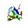
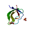
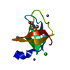
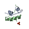
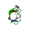
Entry Database : PDB / ID : 5oazTitle Crystal structure of the Abl-SH3 domain at pH 7.5 Tyrosine-protein kinase ABL1 Keywords / Function / homology Function Domain/homology Component
/ / / / / / / / / / / / / / / / / / / / / / / / / / / / / / / / / / / / / / / / / / / / / / / / / / / / / / / / / / / / / / / / / / / / / / / / / / / / / / / / / / / / / / / / / / / / / / / / / / / / / / / / / / / / / / / / / / / / / / / / / / / / / / / / / / / / / / / / / / / / / / / / / / Biological species Homo sapiens (human)Method / / / / Resolution : 1.03 Å Authors Camara-Artigas, A. Funding support Organization Grant number Country Spanish Ministry of Economy and Competitiveness BIO2016-78020-R
Journal : to be published Title : Crystal structure of the Abl-SH3 domain at pH 7.5Authors : Camara-Artigas, A. History Deposition Jun 25, 2017 Deposition site / Processing site Revision 1.0 Jul 12, 2017 Provider / Type Revision 1.1 Sep 13, 2017 Group / Category / Item Revision 1.2 Jan 24, 2018 Group / Category Item / _entity_src_gen.pdbx_host_org_scientific_name / _entity_src_gen.pdbx_host_org_strainRevision 1.3 Jan 17, 2024 Group / Database references / Refinement descriptionCategory chem_comp_atom / chem_comp_bond ... chem_comp_atom / chem_comp_bond / database_2 / pdbx_initial_refinement_model Item / _database_2.pdbx_database_accession
Show all Show less
 Movie
Movie Controller


 Open data
 Basic information
 Components
 Keywords
 Function and homology information
 Homo sapiens (human)
 X-RAY DIFFRACTION /
  Authors
 Spain, 1items
  Citation
 Structure visualization
 Downloads & links
 Other downloads


 PDBj
PDBj


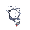
 Assembly


 Mass: 106.120 Da / Num. of mol.: 3 / Source method: obtained synthetically / Formula: C4H10O3 / Feature type: SUBJECT OF INVESTIGATION
 Mass: 18.015 Da / Num. of mol.: 124 / Source method: isolated from a natural source / Formula: H2O
 Sample preparation
 Processing