 Movie
Movie Controller

[English] 日本語
 Yorodumi
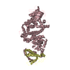
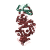
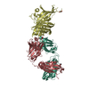
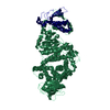
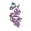
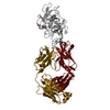
- PDB-7n4l: Crystal structure of SARS-CoV-2 receptor binding domain in comple... -
+ Open data

- Basic information
| Entry | Database: PDB / ID: 7n4l | ||||||
|---|---|---|---|---|---|---|---|
| Title | Crystal structure of SARS-CoV-2 receptor binding domain in complex with neutralizing human antibody WRAIR-2125. | ||||||
 Components Components |
| ||||||
 Keywords Keywords | VIRAL PROTEIN/IMMUNE SYSTEM / Neutralizing antibody / SARS-CoV-2 / WRAIR-2125 / receptor binding domain / ANTIVIRAL PROTEIN / VIRAL PROTEIN-IMMUNE SYSTEM complex | ||||||
| Function / homology |  Function and homology information Function and homology informationsymbiont-mediated disruption of host tissue / Maturation of spike protein / Translation of Structural Proteins / Virion Assembly and Release / host cell surface / host extracellular space / viral translation / symbiont-mediated-mediated suppression of host tetherin activity / Induction of Cell-Cell Fusion / structural constituent of virion ...symbiont-mediated disruption of host tissue / Maturation of spike protein / Translation of Structural Proteins / Virion Assembly and Release / host cell surface / host extracellular space / viral translation / symbiont-mediated-mediated suppression of host tetherin activity / Induction of Cell-Cell Fusion / structural constituent of virion / entry receptor-mediated virion attachment to host cell / membrane fusion / Attachment and Entry / host cell endoplasmic reticulum-Golgi intermediate compartment membrane / positive regulation of viral entry into host cell / receptor-mediated virion attachment to host cell / host cell surface receptor binding / symbiont-mediated suppression of host innate immune response / receptor ligand activity / endocytosis involved in viral entry into host cell / fusion of virus membrane with host plasma membrane / fusion of virus membrane with host endosome membrane / viral envelope / symbiont entry into host cell / virion attachment to host cell / SARS-CoV-2 activates/modulates innate and adaptive immune responses / host cell plasma membrane / virion membrane / identical protein binding / membrane / plasma membrane Similarity search - Function | ||||||
| Biological species |   Severe acute respiratory syndrome coronavirus 2 Severe acute respiratory syndrome coronavirus 2 Homo sapiens (human) Homo sapiens (human) | ||||||
| Method |  X-RAY DIFFRACTION / SYNCHROTRON / MOLECULAR REPLACEMENT / Resolution: 3.601 Å X-RAY DIFFRACTION / SYNCHROTRON / MOLECULAR REPLACEMENT / Resolution: 3.601 Å | ||||||
 Authors Authors | Sankhala, R.S. / Joyce, M.G. | ||||||
| Funding support |  United States, 1items United States, 1items
| ||||||
 Citation Citation | Journal: Nat.Immunol. / Year: 2021 Title: Low-dose in vivo protection and neutralization across SARS-CoV-2 variants by monoclonal antibody combinations. Authors: Dussupt, V. / Sankhala, R.S. / Mendez-Rivera, L. / Townsley, S.M. / Schmidt, F. / Wieczorek, L. / Lal, K.G. / Donofrio, G.C. / Tran, U. / Jackson, N.D. / Zaky, W.I. / Zemil, M. / Tritsch, S. ...Authors: Dussupt, V. / Sankhala, R.S. / Mendez-Rivera, L. / Townsley, S.M. / Schmidt, F. / Wieczorek, L. / Lal, K.G. / Donofrio, G.C. / Tran, U. / Jackson, N.D. / Zaky, W.I. / Zemil, M. / Tritsch, S.R. / Chen, W.H. / Martinez, E.J. / Ahmed, A. / Choe, M. / Chang, W.C. / Hajduczki, A. / Jian, N. / Peterson, C.E. / Rees, P.A. / Rutkowska, M. / Slike, B.M. / Selverian, C.N. / Swafford, I. / Teng, I.T. / Thomas, P.V. / Zhou, T. / Smith, C.J. / Currier, J.R. / Kwong, P.D. / Rolland, M. / Davidson, E. / Doranz, B.J. / Mores, C.N. / Hatziioannou, T. / Reiley, W.W. / Bieniasz, P.D. / Paquin-Proulx, D. / Gromowski, G.D. / Polonis, V.R. / Michael, N.L. / Modjarrad, K. / Joyce, M.G. / Krebs, S.J. | ||||||
| History |
|
- Structure visualization
| Structure viewer | Molecule: MolmilJmol/JSmol |
|---|
- Downloads & links
-Download
| PDBx/mmCIF format | 7n4l.cif.gz | 136.3 KB | Display | PDBx/mmCIF format |
|---|---|---|---|---|
| PDB format | pdb7n4l.ent.gz | 104.4 KB | Display | PDB format |
| PDBx/mmJSON format | 7n4l.json.gz | Tree view | PDBx/mmJSON format | |
| Others |  Other downloads Other downloads |
-Validation report
| Summary document | 7n4l_validation.pdf.gz | 444 KB | Display | wwPDB validaton report |
|---|---|---|---|---|
| Full document | 7n4l_full_validation.pdf.gz | 455.2 KB | Display | |
| Data in XML | 7n4l_validation.xml.gz | 23.9 KB | Display | |
| Data in CIF | 7n4l_validation.cif.gz | 32.2 KB | Display | |
| Arichive directory | https://data.pdbj.org/pub/pdb/validation_reports/n4/7n4lftp://data.pdbj.org/pub/pdb/validation_reports/n4/7n4l | HTTPS FTP |
-Related structure data
| Related structure data | 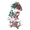 7n4iC  7n4jC 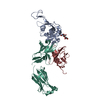 7n4mC 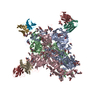 7l2eS S: Starting model for refinement C: citing same article ( |
|---|---|
| Similar structure data |


-Links
 PDBj
PDBj


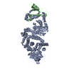
- Assembly
| Deposited unit | 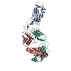
| ||||||||
|---|---|---|---|---|---|---|---|---|---|
| 1 |
| ||||||||
| Unit cell |
|
-Components
| #1: Protein | Mass: 23073.766 Da / Num. of mol.: 1 / Fragment: receptor binding domain (RBD) Source method: isolated from a genetically manipulated source Source: (gene. exp.) Severe acute respiratory syndrome coronavirus 2Gene: S, 2 / Production host: Homo sapiens (human) / References: UniProt: P0DTC2 |
|---|---|
| #2: Antibody | Mass: 24842.635 Da / Num. of mol.: 1 Source method: isolated from a genetically manipulated source Source: (gene. exp.) Homo sapiens (human) / Production host: Homo sapiens (human) |
| #3: Antibody | Mass: 23424.939 Da / Num. of mol.: 1 Source method: isolated from a genetically manipulated source Source: (gene. exp.) Homo sapiens (human) / Production host: Homo sapiens (human) |
| Has protein modification | Y |
-Experimental details
-Experiment
| Experiment | Method: X-RAY DIFFRACTION / Number of used crystals: 1 |
|---|
- Sample preparation
| Crystal | Density Matthews: 4.7 Å3/Da / Density % sol: 73.85 % |
|---|---|
| Crystal grow | Temperature: 291 K / Method: vapor diffusion, hanging drop / pH: 8.5 Details: 0.12 M alcohol mixture (1,6-Hexanediol; 1-Butanol; 1,2-Propanediol; 2-Propanol; 1,4-Butanediol; 1,3-Propanediol), 0.1M buffer system 3 (Tris base and BICINE, pH 8.5), 50% precipitant mix 4 ...Details: 0.12 M alcohol mixture (1,6-Hexanediol; 1-Butanol; 1,2-Propanediol; 2-Propanol; 1,4-Butanediol; 1,3-Propanediol), 0.1M buffer system 3 (Tris base and BICINE, pH 8.5), 50% precipitant mix 4 (25% v/v MPD; 25% PEG 1000; 25% w/v PEG 3350) and 0.1 M Manganese(II) chloride tetrahydrate |
-Data collection
| Diffraction | Mean temperature: 100 K / Serial crystal experiment: N |
|---|---|
| Diffraction source | Source: SYNCHROTRON / Site: APS / Beamline: 21-ID-E / Wavelength: 0.97 Å |
| Detector | Type: DECTRIS EIGER X 16M / Detector: PIXEL / Date: Apr 21, 2021 |
| Radiation | Protocol: SINGLE WAVELENGTH / Monochromatic (M) / Laue (L): M / Scattering type: x-ray |
| Radiation wavelength | Wavelength: 0.97 Å / Relative weight: 1 |
| Reflection | Resolution: 3.43→163.6 Å / Num. obs: 16510 / % possible obs: 78 % / Redundancy: 2.4 % / CC1/2: 0.98 / Net I/σ(I): 4.3 |
| Reflection shell | Resolution: 3.43→3.71 Å / Num. unique obs: 8436 / CC1/2: 0.4 |
- Processing
| Software |
| ||||||||||||||||||||||||||||||
|---|---|---|---|---|---|---|---|---|---|---|---|---|---|---|---|---|---|---|---|---|---|---|---|---|---|---|---|---|---|---|---|
| Refinement | Method to determine structure: MOLECULAR REPLACEMENT Starting model: 7l2e Resolution: 3.601→19.983 Å / SU ML: 0.6 / Cross valid method: THROUGHOUT / σ(F): 1.36 / Phase error: 33.89 / Stereochemistry target values: ML
| ||||||||||||||||||||||||||||||
| Solvent computation | Shrinkage radii: 0.9 Å / VDW probe radii: 1.11 Å / Solvent model: FLAT BULK SOLVENT MODEL | ||||||||||||||||||||||||||||||
| Displacement parameters | Biso max: 126.76 Å2 / Biso mean: 41.1011 Å2 / Biso min: 12.05 Å2 | ||||||||||||||||||||||||||||||
| Refinement step | Cycle: final / Resolution: 3.601→19.983 Å
| ||||||||||||||||||||||||||||||
| Refine LS restraints |
| ||||||||||||||||||||||||||||||
| LS refinement shell | Refine-ID: X-RAY DIFFRACTION / Rfactor Rfree error: 0
|
