 Movie
Movie Controller

[English] 日本語
 Yorodumi
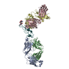
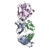
- PDB-6xzw: Crystal structure of the meningococcal vaccine antigen fHbp in co... -
+ Open data

- Basic information
| Entry | Database: PDB / ID: 6xzw | ||||||
|---|---|---|---|---|---|---|---|
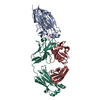
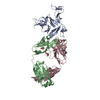
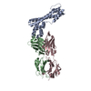
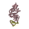
| Title | Crystal structure of the meningococcal vaccine antigen fHbp in complex with a cross-reactive human Fab. | ||||||
 Components Components |
| ||||||
 Keywords Keywords | IMMUNE SYSTEM / Antigen-antibody complex / epitope mapping / lipoprotein / neisseria meningitidis | ||||||
| Function / homology |  Function and homology information Function and homology information | ||||||
| Biological species |  Neisseria meningitidis serogroup B (bacteria) Neisseria meningitidis serogroup B (bacteria) Homo sapiens (human) Homo sapiens (human) | ||||||
| Method |  X-RAY DIFFRACTION / SYNCHROTRON / MOLECULAR REPLACEMENT / Resolution: 2.4 Å X-RAY DIFFRACTION / SYNCHROTRON / MOLECULAR REPLACEMENT / Resolution: 2.4 Å | ||||||
 Authors Authors | Veggi, D. / Cozzi, R. | ||||||
 Citation Citation | Journal: Plos Pathog. / Year: 2020 Title: 4CMenB vaccine induces elite cross-protective human antibodies that compete with human factor H for binding to meningococcal fHbp. Authors: Veggi, D. / Bianchi, F. / Santini, L. / Lo Surdo, P. / Chesterman, C.C. / Pansegrau, W. / Bechi, N. / Huang, Y. / Masignani, V. / Pizza, M. / Rappuoli, R. / Bottomley, M.J. / Cozzi, R. / Maione, D. | ||||||
| History |
|
- Structure visualization
| Structure viewer | Molecule: MolmilJmol/JSmol |
|---|
- Downloads & links
-Download
| PDBx/mmCIF format | 6xzw.cif.gz | 155 KB | Display | PDBx/mmCIF format |
|---|---|---|---|---|
| PDB format | pdb6xzw.ent.gz | 109.2 KB | Display | PDB format |
| PDBx/mmJSON format | 6xzw.json.gz | Tree view | PDBx/mmJSON format | |
| Others |  Other downloads Other downloads |
-Validation report
| Arichive directory | https://data.pdbj.org/pub/pdb/validation_reports/xz/6xzwftp://data.pdbj.org/pub/pdb/validation_reports/xz/6xzw | HTTPS FTP |
|---|
-Related structure data


-Links
 PDBj
PDBj


- Assembly
| Deposited unit | 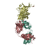
| ||||||||||||
|---|---|---|---|---|---|---|---|---|---|---|---|---|---|
| 1 |
| ||||||||||||
| Unit cell |
|
-Components
| #1: Protein | Mass: 26896.045 Da / Num. of mol.: 1 Source method: isolated from a genetically manipulated source Source: (gene. exp.) Neisseria meningitidis serogroup B (strain MC58) (bacteria)Gene: NMB1870 / Production host: | ||||||
|---|---|---|---|---|---|---|---|
| #2: Antibody | Mass: 23787.438 Da / Num. of mol.: 1 Source method: isolated from a genetically manipulated source Source: (gene. exp.) Homo sapiens (human) / Production host: Homo sapiens (human) | ||||||
| #3: Antibody | Mass: 23642.670 Da / Num. of mol.: 1 Source method: isolated from a genetically manipulated source Source: (gene. exp.) Homo sapiens (human) / Production host: Homo sapiens (human) | ||||||
| #4: Chemical |   Mass: 62.068 Da / Num. of mol.: 3 / Source method: obtained synthetically / Formula: C2H6O2 Mass: 62.068 Da / Num. of mol.: 3 / Source method: obtained synthetically / Formula: C2H6O2#5: Water | ChemComp-HOH / |  Mass: 18.015 Da / Num. of mol.: 56 / Source method: isolated from a natural source / Formula: H2O Mass: 18.015 Da / Num. of mol.: 56 / Source method: isolated from a natural source / Formula: H2OHas ligand of interest | N | Has protein modification | Y | |
-Experimental details
-Experiment
| Experiment | Method: X-RAY DIFFRACTION / Number of used crystals: 1 |
|---|
- Sample preparation
| Crystal | Density Matthews: 2.79 Å3/Da / Density % sol: 55.85 % |
|---|---|
| Crystal grow | Temperature: 293.15 K / Method: vapor diffusion, sitting drop / Details: 0.1M HEPES, 20% w/v jeff ED-2001, pH 6.5 |
-Data collection
| Diffraction | Mean temperature: 100 K / Serial crystal experiment: N |
|---|---|
| Diffraction source | Source: SYNCHROTRON / Site: ESRF  / Beamline: ID29 / Wavelength: 0.966 Å / Beamline: ID29 / Wavelength: 0.966 Å |
| Detector | Type: DECTRIS PILATUS 2M / Detector: PIXEL / Date: Jul 17, 2017 |
| Radiation | Protocol: SINGLE WAVELENGTH / Monochromatic (M) / Laue (L): M / Scattering type: x-ray |
| Radiation wavelength | Wavelength: 0.966 Å / Relative weight: 1 |
| Reflection | Resolution: 2.4→46.78 Å / Num. obs: 29802 / % possible obs: 93 % / Redundancy: 3.4 % / Biso Wilson estimate: 50.17 Å2 / CC1/2: 0.99 / Net I/σ(I): 16.5 |
| Reflection shell | Resolution: 2.4→2.48 Å / Num. unique obs: 3202 / CC1/2: 0.82 |
- Processing
| Software |
| ||||||||||||||||||
|---|---|---|---|---|---|---|---|---|---|---|---|---|---|---|---|---|---|---|---|
| Refinement | Method to determine structure: MOLECULAR REPLACEMENT Starting model: 3KVD, 5TLK Resolution: 2.4→46.78 Å / SU ML: 0.31 / Cross valid method: FREE R-VALUE / σ(F): 1.34 / Phase error: 29.48
| ||||||||||||||||||
| Solvent computation | Shrinkage radii: 0.9 Å / VDW probe radii: 1.11 Å | ||||||||||||||||||
| Displacement parameters | Biso mean: 55.68 Å2 | ||||||||||||||||||
| Refinement step | Cycle: LAST / Resolution: 2.4→46.78 Å
| ||||||||||||||||||
| Refine LS restraints | Type: f_chiral_restr / Dev ideal: 0.0735652181087 / Number: 779 | ||||||||||||||||||
| LS refinement shell | Resolution: 2.4→2.48 Å
|
