 Movie
Movie Controller

[English] 日本語
 Yorodumi
- PDB-7myz: Structure of the full length 5-TM receptor CD47 bound to Fab B6H12 -
+ Open data

- Basic information
| Entry | Database: PDB / ID: 7myz | ||||||
|---|---|---|---|---|---|---|---|
| Title | Structure of the full length 5-TM receptor CD47 bound to Fab B6H12 | ||||||
 Components Components |
| ||||||
 Keywords Keywords | MEMBRANE PROTEIN / 5-TM receptor / immune receptor / phagocytosis inhibitor | ||||||
| Function / homology |  Function and homology information Function and homology informationcellular response to interleukin-12 / regulation of Fc receptor mediated stimulatory signaling pathway / protein binding involved in heterotypic cell-cell adhesion / positive regulation of monocyte extravasation / regulation of interleukin-10 production / cell-cell adhesion mediator activity / positive regulation of cell-cell adhesion / regulation of type II interferon production / ATP export / fibrinogen binding ...cellular response to interleukin-12 / regulation of Fc receptor mediated stimulatory signaling pathway / protein binding involved in heterotypic cell-cell adhesion / positive regulation of monocyte extravasation / regulation of interleukin-10 production / cell-cell adhesion mediator activity / positive regulation of cell-cell adhesion / regulation of type II interferon production / ATP export / fibrinogen binding / regulation of tumor necrosis factor production / regulation of interleukin-12 production / regulation of nitric oxide biosynthetic process / regulation of interleukin-6 production / Signal regulatory protein family interactions / negative regulation of phagocytosis / thrombospondin receptor activity / tertiary granule membrane / cellular response to interleukin-1 / Integrin cell surface interactions / specific granule membrane / positive regulation of stress fiber assembly / positive regulation of phagocytosis / Cell surface interactions at the vascular wall / integrin-mediated signaling pathway / electron transport chain / cellular response to type II interferon / positive regulation of T cell activation / positive regulation of inflammatory response / cell migration / angiogenesis / electron transfer activity / periplasmic space / iron ion binding / inflammatory response / apoptotic process / heme binding / positive regulation of cell population proliferation / Neutrophil degranulation / cell surface / extracellular exosome / plasma membrane Similarity search - Function | ||||||
| Biological species |  Homo sapiens (human) Homo sapiens (human) | ||||||
| Method |  X-RAY DIFFRACTION / SYNCHROTRON / MOLECULAR REPLACEMENT / Resolution: 3.4 Å X-RAY DIFFRACTION / SYNCHROTRON / MOLECULAR REPLACEMENT / Resolution: 3.4 Å | ||||||
 Authors Authors | Fenalti, G. / Villanueva, N. | ||||||
 Citation Citation | Journal: Nat Commun / Year: 2021 Title: Structure of the human marker of self 5-transmembrane receptor CD47. Authors: Fenalti, G. / Villanueva, N. / Griffith, M. / Pagarigan, B. / Lakkaraju, S.K. / Huang, R.Y. / Ladygina, N. / Sharma, A. / Mikolon, D. / Abbasian, M. / Johnson, J. / Hadjivassiliou, H. / Zhu, ...Authors: Fenalti, G. / Villanueva, N. / Griffith, M. / Pagarigan, B. / Lakkaraju, S.K. / Huang, R.Y. / Ladygina, N. / Sharma, A. / Mikolon, D. / Abbasian, M. / Johnson, J. / Hadjivassiliou, H. / Zhu, D. / Chamberlain, P.P. / Cho, H. / Hariharan, K. | ||||||
| History |
|
- Structure visualization
| Structure viewer | Molecule: MolmilJmol/JSmol |
|---|
- Downloads & links
-Download
| PDBx/mmCIF format | 7myz.cif.gz | 255.1 KB | Display | PDBx/mmCIF format |
|---|---|---|---|---|
| PDB format | pdb7myz.ent.gz | 196.2 KB | Display | PDB format |
| PDBx/mmJSON format | 7myz.json.gz | Tree view | PDBx/mmJSON format | |
| Others |  Other downloads Other downloads |
-Validation report
| Arichive directory | https://data.pdbj.org/pub/pdb/validation_reports/my/7myzftp://data.pdbj.org/pub/pdb/validation_reports/my/7myz | HTTPS FTP |
|---|
-Related structure data
| Related structure data | 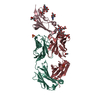 5tzuS S: Starting model for refinement |
|---|---|
| Similar structure data |


-Links
 PDBj
PDBj


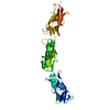
- Assembly
| Deposited unit | 
| |||||||||||||||||||||||||||||||||||||||||||||||||||||||||||||||||||||||||||||||||||||||||||||||
|---|---|---|---|---|---|---|---|---|---|---|---|---|---|---|---|---|---|---|---|---|---|---|---|---|---|---|---|---|---|---|---|---|---|---|---|---|---|---|---|---|---|---|---|---|---|---|---|---|---|---|---|---|---|---|---|---|---|---|---|---|---|---|---|---|---|---|---|---|---|---|---|---|---|---|---|---|---|---|---|---|---|---|---|---|---|---|---|---|---|---|---|---|---|---|---|---|
| 1 | 
| |||||||||||||||||||||||||||||||||||||||||||||||||||||||||||||||||||||||||||||||||||||||||||||||
| 2 | 
| |||||||||||||||||||||||||||||||||||||||||||||||||||||||||||||||||||||||||||||||||||||||||||||||
| Unit cell |
| |||||||||||||||||||||||||||||||||||||||||||||||||||||||||||||||||||||||||||||||||||||||||||||||
| Noncrystallographic symmetry (NCS) | NCS domain:
NCS domain segments: Component-ID: _ / Refine code: _
NCS ensembles :
|
-Components
-Antibody , 3 types, 6 molecules CDHILM
| #1: Antibody | Mass: 46644.930 Da / Num. of mol.: 2 Source method: isolated from a genetically manipulated source Source: (gene. exp.) Homo sapiens (human), (gene. exp.) Gene: CD47, MER6, cybC / Production host:   Spodoptera frugiperda (fall armyworm) / References: UniProt: Q08722, UniProt: P0ABE7 Spodoptera frugiperda (fall armyworm) / References: UniProt: Q08722, UniProt: P0ABE7#2: Antibody | Mass: 23264.068 Da / Num. of mol.: 2 Source method: isolated from a genetically manipulated source Source: (gene. exp.) Homo sapiens (human) / Production host:   Cricetulus griseus (Chinese hamster) Cricetulus griseus (Chinese hamster)#3: Antibody | Mass: 23270.766 Da / Num. of mol.: 2 Source method: isolated from a genetically manipulated source Source: (gene. exp.) Homo sapiens (human) / Production host: Cricetulus griseus (Chinese hamster) |
|---|
-Sugars , 1 types, 7 molecules 
| #4: Sugar | ChemComp-NAG /  Type: D-saccharide, beta linking / Mass: 221.208 Da / Num. of mol.: 7 Type: D-saccharide, beta linking / Mass: 221.208 Da / Num. of mol.: 7Source method: isolated from a genetically manipulated source Formula: C8H15NO6 |
|---|
-Non-polymers , 2 types, 4 molecules 

| #5: Chemical | ChemComp-GOL /  Mass: 92.094 Da / Num. of mol.: 1 / Source method: obtained synthetically / Formula: C3H8O3 Mass: 92.094 Da / Num. of mol.: 1 / Source method: obtained synthetically / Formula: C3H8O3 |
|---|---|
| #6: Water | ChemComp-HOH / Mass: 18.015 Da / Num. of mol.: 3 / Source method: isolated from a natural source / Formula: H2O |
-Details
| Has ligand of interest | N |
|---|---|
| Has protein modification | Y |
-Experimental details
-Experiment
| Experiment | Method: X-RAY DIFFRACTION / Number of used crystals: 1 |
|---|
- Sample preparation
| Crystal | Density Matthews: 3.7 Å3/Da / Density % sol: 66.78 % |
|---|---|
| Crystal grow | Temperature: 293 K / Method: lipidic cubic phase / pH: 6 Details: 100mM sodium citrate pH 6.0, 450mM ammonium acetate, 32% PEG400 PH range: 5.9-6.2 |
-Data collection
| Diffraction | Mean temperature: 100 K / Serial crystal experiment: N | ||||||||||||||||||||||||||||||||||||||||||||||||||||||||||||||||||||||||||||||||||||||||||||||||||||||||||||||||||||||||||||||||||||||||||||||||
|---|---|---|---|---|---|---|---|---|---|---|---|---|---|---|---|---|---|---|---|---|---|---|---|---|---|---|---|---|---|---|---|---|---|---|---|---|---|---|---|---|---|---|---|---|---|---|---|---|---|---|---|---|---|---|---|---|---|---|---|---|---|---|---|---|---|---|---|---|---|---|---|---|---|---|---|---|---|---|---|---|---|---|---|---|---|---|---|---|---|---|---|---|---|---|---|---|---|---|---|---|---|---|---|---|---|---|---|---|---|---|---|---|---|---|---|---|---|---|---|---|---|---|---|---|---|---|---|---|---|---|---|---|---|---|---|---|---|---|---|---|---|---|---|---|---|
| Diffraction source | Source: SYNCHROTRON / Site: Diamond  / Beamline: I24 / Wavelength: 0.987 Å / Beamline: I24 / Wavelength: 0.987 Å | ||||||||||||||||||||||||||||||||||||||||||||||||||||||||||||||||||||||||||||||||||||||||||||||||||||||||||||||||||||||||||||||||||||||||||||||||
| Detector | Type: DECTRIS PILATUS 6M / Detector: PIXEL / Date: Feb 24, 2020 | ||||||||||||||||||||||||||||||||||||||||||||||||||||||||||||||||||||||||||||||||||||||||||||||||||||||||||||||||||||||||||||||||||||||||||||||||
| Radiation | Protocol: SINGLE WAVELENGTH / Monochromatic (M) / Laue (L): M / Scattering type: x-ray | ||||||||||||||||||||||||||||||||||||||||||||||||||||||||||||||||||||||||||||||||||||||||||||||||||||||||||||||||||||||||||||||||||||||||||||||||
| Radiation wavelength | Wavelength: 0.987 Å / Relative weight: 1 | ||||||||||||||||||||||||||||||||||||||||||||||||||||||||||||||||||||||||||||||||||||||||||||||||||||||||||||||||||||||||||||||||||||||||||||||||
| Reflection | Resolution: 3.4→50 Å / Num. obs: 35403 / % possible obs: 92.1 % / Redundancy: 3.3 % / Rmerge(I) obs: 0.358 / Rpim(I) all: 0.206 / Rrim(I) all: 0.417 / Χ2: 1.389 / Net I/σ(I): 3.7 / Num. measured all: 118336 | ||||||||||||||||||||||||||||||||||||||||||||||||||||||||||||||||||||||||||||||||||||||||||||||||||||||||||||||||||||||||||||||||||||||||||||||||
| Reflection shell | Diffraction-ID: 1
|
- Processing
| Software |
| ||||||||||||||||||||||||||||||||||||||||||||||||||||||||||||
|---|---|---|---|---|---|---|---|---|---|---|---|---|---|---|---|---|---|---|---|---|---|---|---|---|---|---|---|---|---|---|---|---|---|---|---|---|---|---|---|---|---|---|---|---|---|---|---|---|---|---|---|---|---|---|---|---|---|---|---|---|---|
| Refinement | Method to determine structure: MOLECULAR REPLACEMENT Starting model: 5TZU Resolution: 3.4→48.68 Å / Cor.coef. Fo:Fc: 0.86 / Cor.coef. Fo:Fc free: 0.841 / SU B: 28.196 / SU ML: 0.424 / Cross valid method: THROUGHOUT / σ(F): 0 / ESU R Free: 0.516 / Stereochemistry target values: MAXIMUM LIKELIHOOD Details: HYDROGENS HAVE BEEN ADDED IN THE RIDING POSITIONS U VALUES : REFINED INDIVIDUALLY
| ||||||||||||||||||||||||||||||||||||||||||||||||||||||||||||
| Solvent computation | Ion probe radii: 0.8 Å / Shrinkage radii: 0.8 Å / VDW probe radii: 1.2 Å / Solvent model: MASK | ||||||||||||||||||||||||||||||||||||||||||||||||||||||||||||
| Displacement parameters | Biso max: 248.36 Å2 / Biso mean: 78.056 Å2 / Biso min: 13.28 Å2
| ||||||||||||||||||||||||||||||||||||||||||||||||||||||||||||
| Refinement step | Cycle: final / Resolution: 3.4→48.68 Å
| ||||||||||||||||||||||||||||||||||||||||||||||||||||||||||||
| Refine LS restraints |
| ||||||||||||||||||||||||||||||||||||||||||||||||||||||||||||
| Refine LS restraints NCS | Refine-ID: X-RAY DIFFRACTION / Type: interatomic distance / Weight position: 0.05
| ||||||||||||||||||||||||||||||||||||||||||||||||||||||||||||
| LS refinement shell | Resolution: 3.4→3.488 Å / Rfactor Rfree error: 0 / Total num. of bins used: 20
|
