 Movie
Movie Controller

[English] 日本語
 Yorodumi
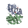
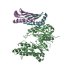
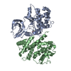
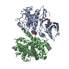
- PDB-7mtl: Crystal structure of colibactin self-resistance protein ClbS in c... -
+ Open data

- Basic information
| Entry | Database: PDB / ID: 7mtl | ||||||
|---|---|---|---|---|---|---|---|
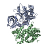
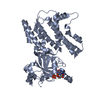
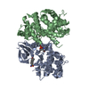
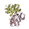
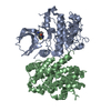
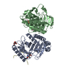
| Title | Crystal structure of colibactin self-resistance protein ClbS in complex with a dsDNA | ||||||
 Components Components |
| ||||||
 Keywords Keywords | DNA BINDING PROTEIN/DNA / hydrolase / DNA repair / DNA BINDING PROTEIN-DNA complex | ||||||
| Function / homology | Protein of unknown function DUF1706 / Protein of unknown function (DUF1706) / DinB/YfiT-like putative metalloenzymes / DNA / DNA (> 10) / Colibactin self-protection protein ClbS Function and homology information Function and homology information | ||||||
| Biological species |  Synthetic construct (others) | ||||||
| Method |  X-RAY DIFFRACTION / SYNCHROTRON / MOLECULAR REPLACEMENT / Resolution: 2.446 Å X-RAY DIFFRACTION / SYNCHROTRON / MOLECULAR REPLACEMENT / Resolution: 2.446 Å | ||||||
 Authors Authors | Tripathi, P. / Bruner, S.D. | ||||||
 Citation Citation | Journal: Biochemistry / Year: 2021 Title: Structural Basis for the Interactions of the Colibactin Resistance Gene Product ClbS with DNA. Authors: Tripathi, P. / Bruner, S.D. #1: Journal: Journal of American Chemical Society / Year: 2017Title: ClbS Is a Cyclopropane Hydrolase That Confers Colibactin Resistance Authors: Tripathi, P. / Bruner, S.D. | ||||||
| History |
|
- Structure visualization
| Structure viewer | Molecule: MolmilJmol/JSmol |
|---|
- Downloads & links
-Download
| PDBx/mmCIF format | 7mtl.cif.gz | 98.8 KB | Display | PDBx/mmCIF format |
|---|---|---|---|---|
| PDB format | pdb7mtl.ent.gz | 71.6 KB | Display | PDB format |
| PDBx/mmJSON format | 7mtl.json.gz | Tree view | PDBx/mmJSON format | |
| Others |  Other downloads Other downloads |
-Validation report
| Summary document | 7mtl_validation.pdf.gz | 444.6 KB | Display | wwPDB validaton report |
|---|---|---|---|---|
| Full document | 7mtl_full_validation.pdf.gz | 448.5 KB | Display | |
| Data in XML | 7mtl_validation.xml.gz | 14.4 KB | Display | |
| Data in CIF | 7mtl_validation.cif.gz | 19.1 KB | Display | |
| Arichive directory | https://data.pdbj.org/pub/pdb/validation_reports/mt/7mtlftp://data.pdbj.org/pub/pdb/validation_reports/mt/7mtl | HTTPS FTP |
-Related structure data
| Related structure data | 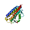 7mttC 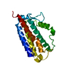 6anrS S: Starting model for refinement C: citing same article ( |
|---|---|
| Similar structure data |
-Links
 PDBj
PDBj


- Assembly
| Deposited unit | 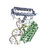
| ||||||||
|---|---|---|---|---|---|---|---|---|---|
| 1 |
| ||||||||
| Unit cell |
|
-Components
| #1: Protein | Mass: 20747.664 Da / Num. of mol.: 2 Source method: isolated from a genetically manipulated source Source: (gene. exp.) Gene: clbS, APU18_08100, AWF59_018730, AZZ83_004503, CT146_21495, CUB99_02165, D3C88_24730, DNR41_00045, DNX30_29170, DS966_21615, DU333_02935, DW236_02620, ELT23_23590, ELT33_24270, EPS70_02685, ...Gene: clbS, APU18_08100, AWF59_018730, AZZ83_004503, CT146_21495, CUB99_02165, D3C88_24730, DNR41_00045, DNX30_29170, DS966_21615, DU333_02935, DW236_02620, ELT23_23590, ELT33_24270, EPS70_02685, EPS94_00180, EWK56_23755, FPI65_12320, FQU83_11985, GFU47_04510, GP945_05010, GP946_20500, HHG54_004716, HHJ44_00090, HJL93_000008, HJM41_001166, HJO44_004707, HJS53_002303, HmCmsJML146_00176, HNX34_25610, HV055_09710, HV098_09980, HV348_09415, NCTC9075_02752, NCTC9434_01964 Production host: #2: DNA chain | | Mass: 4488.938 Da / Num. of mol.: 1 / Source method: obtained synthetically / Source: (synth.) Synthetic construct (others) #3: DNA chain | | Mass: 4689.057 Da / Num. of mol.: 1 / Source method: obtained synthetically / Source: (synth.) Synthetic construct (others) #4: Water | ChemComp-HOH / |  Mass: 18.015 Da / Num. of mol.: 10 / Source method: isolated from a natural source / Formula: H2O Mass: 18.015 Da / Num. of mol.: 10 / Source method: isolated from a natural source / Formula: H2O |
|---|
-Experimental details
-Experiment
| Experiment | Method: X-RAY DIFFRACTION / Number of used crystals: 1 |
|---|
- Sample preparation
| Crystal | Density Matthews: 2.98 Å3/Da / Density % sol: 58.75 % |
|---|---|
| Crystal grow | Temperature: 277 K / Method: vapor diffusion, sitting drop Details: 0.2 M sodium acetate trihydrate pH 7.0, 20% (w/v) PEG 3350 |
-Data collection
| Diffraction | Mean temperature: 100 K / Serial crystal experiment: N |
|---|---|
| Diffraction source | Source: SYNCHROTRON / Site: APS  / Beamline: 21-ID-G / Wavelength: 0.9787 Å / Beamline: 21-ID-G / Wavelength: 0.9787 Å |
| Detector | Type: MARMOSAIC 300 mm CCD / Detector: CCD / Date: Dec 5, 2019 |
| Radiation | Protocol: SINGLE WAVELENGTH / Monochromatic (M) / Laue (L): M / Scattering type: x-ray |
| Radiation wavelength | Wavelength: 0.9787 Å / Relative weight: 1 |
| Reflection | Resolution: 2.446→46.793 Å / Num. obs: 22255 / % possible obs: 99.86 % / Redundancy: 6.6 % / CC1/2: 0.998 / Net I/σ(I): 11.79 |
| Reflection shell | Resolution: 2.446→2.533 Å / Mean I/σ(I) obs: 1.44 / Num. unique obs: 2168 / CC1/2: 0.755 / % possible all: 98.77 |
- Processing
| Software |
| ||||||||||||||||||||||||||||||||||||||||||||||||||||||
|---|---|---|---|---|---|---|---|---|---|---|---|---|---|---|---|---|---|---|---|---|---|---|---|---|---|---|---|---|---|---|---|---|---|---|---|---|---|---|---|---|---|---|---|---|---|---|---|---|---|---|---|---|---|---|---|
| Refinement | Method to determine structure: MOLECULAR REPLACEMENT Starting model: 6ANR Resolution: 2.446→46.793 Å / SU ML: 0.4 / Cross valid method: FREE R-VALUE / σ(F): 1.34 / Phase error: 35.11 / Stereochemistry target values: ML
| ||||||||||||||||||||||||||||||||||||||||||||||||||||||
| Solvent computation | Shrinkage radii: 0.9 Å / VDW probe radii: 1.11 Å / Solvent model: FLAT BULK SOLVENT MODEL | ||||||||||||||||||||||||||||||||||||||||||||||||||||||
| Displacement parameters | Biso max: 140.75 Å2 / Biso mean: 64.8658 Å2 / Biso min: 29.3 Å2 | ||||||||||||||||||||||||||||||||||||||||||||||||||||||
| Refinement step | Cycle: final / Resolution: 2.446→46.793 Å
| ||||||||||||||||||||||||||||||||||||||||||||||||||||||
| LS refinement shell | Refine-ID: X-RAY DIFFRACTION / Rfactor Rfree error: 0
|
