 Movie
Movie Controller

[English] 日本語
 Yorodumi
- PDB-3eno: Crystal structure of Pyrococcus furiosus Pcc1 in complex with The... -
+ Open data

- Basic information
| Entry | Database: PDB / ID: 3eno | ||||||
|---|---|---|---|---|---|---|---|
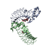
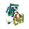
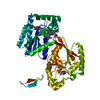
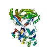
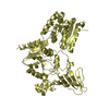
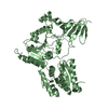
| Title | Crystal structure of Pyrococcus furiosus Pcc1 in complex with Thermoplasma acidophilum Kae1 | ||||||
 Components Components |
| ||||||
 Keywords Keywords | hydrolase/unknown function / Hydrolase / Metal-binding / Metalloprotease / Protease / Zinc / KEOPS complex / ATPase / metal ion binding / dimerization module / telomere / hydrolase-unknown function COMPLEX | ||||||
| Function / homology |  Function and homology information Function and homology informationN6-L-threonylcarbamoyladenine synthase / tRNA N(6)-L-threonylcarbamoyladenine synthase activity / EKC/KEOPS complex / tRNA threonylcarbamoyladenosine modification / protein serine/threonine/tyrosine kinase activity / metalloendopeptidase activity / non-specific serine/threonine protein kinase / iron ion binding / protein serine kinase activity / protein serine/threonine kinase activity ...N6-L-threonylcarbamoyladenine synthase / tRNA N(6)-L-threonylcarbamoyladenine synthase activity / EKC/KEOPS complex / tRNA threonylcarbamoyladenosine modification / protein serine/threonine/tyrosine kinase activity / metalloendopeptidase activity / non-specific serine/threonine protein kinase / iron ion binding / protein serine kinase activity / protein serine/threonine kinase activity / zinc ion binding / ATP binding / cytoplasm Similarity search - Function | ||||||
| Biological species |   Thermoplasma acidophilum (acidophilic)Pyrococcus furiosus (archaea) Thermoplasma acidophilum (acidophilic)Pyrococcus furiosus (archaea) | ||||||
| Method |  X-RAY DIFFRACTION / SYNCHROTRON / MOLECULAR REPLACEMENT / Resolution: 3.0201 Å X-RAY DIFFRACTION / SYNCHROTRON / MOLECULAR REPLACEMENT / Resolution: 3.0201 Å | ||||||
 Authors Authors | Neculai, D. | ||||||
 Citation Citation | Journal: Mol.Cell / Year: 2008 Title: Atomic structure of the KEOPS complex: an ancient protein kinase-containing molecular machine. Authors: Mao, D.Y. / Neculai, D. / Downey, M. / Orlicky, S. / Haffani, Y.Z. / Ceccarelli, D.F. / Ho, J.S. / Szilard, R.K. / Zhang, W. / Ho, C.S. / Wan, L. / Fares, C. / Rumpel, S. / Kurinov, I. / ...Authors: Mao, D.Y. / Neculai, D. / Downey, M. / Orlicky, S. / Haffani, Y.Z. / Ceccarelli, D.F. / Ho, J.S. / Szilard, R.K. / Zhang, W. / Ho, C.S. / Wan, L. / Fares, C. / Rumpel, S. / Kurinov, I. / Arrowsmith, C.H. / Durocher, D. / Sicheri, F. | ||||||
| History |
|
- Structure visualization
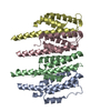
| Structure viewer | Molecule: MolmilJmol/JSmol |
|---|
- Downloads & links
-Download
| PDBx/mmCIF format | 3eno.cif.gz | 194.6 KB | Display | PDBx/mmCIF format |
|---|---|---|---|---|
| PDB format | pdb3eno.ent.gz | 154.9 KB | Display | PDB format |
| PDBx/mmJSON format | 3eno.json.gz | Tree view | PDBx/mmJSON format | |
| Others |  Other downloads Other downloads |
-Validation report
| Arichive directory | https://data.pdbj.org/pub/pdb/validation_reports/en/3enoftp://data.pdbj.org/pub/pdb/validation_reports/en/3eno | HTTPS FTP |
|---|
-Related structure data
| Related structure data | 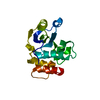 2k8yC 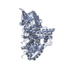 3en9C 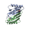 3encC 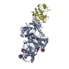 3enhC  3enpC  2ivpS C: citing same article ( S: Starting model for refinement |
|---|---|
| Similar structure data |
-Links
 PDBj
PDBj
- Assembly
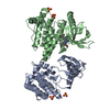
| Deposited unit | 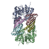
| ||||||||
|---|---|---|---|---|---|---|---|---|---|
| 1 | 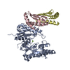
| ||||||||
| 2 | 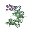
| ||||||||
| Unit cell |
|
-Components
| #1: Protein | Mass: 36056.770 Da / Num. of mol.: 2 Source method: isolated from a genetically manipulated source Source: (gene. exp.) Thermoplasma acidophilum (acidophilic) / Strain: DSM 1728 / Gene: gcp, TA0324 / Plasmid: pGEX / Production host:  #2: Protein | Mass: 9900.515 Da / Num. of mol.: 4 / Mutation: I12M Source method: isolated from a genetically manipulated source Source: (gene. exp.) Pyrococcus furiosus (archaea) / Strain: DSM 3638 / Gene: PF2011 / Plasmid: pGEX / Production host: #3: Chemical |   Mass: 24.305 Da / Num. of mol.: 2 / Source method: obtained synthetically / Formula: Mg Mass: 24.305 Da / Num. of mol.: 2 / Source method: obtained synthetically / Formula: Mg |
|---|
-Experimental details
-Experiment
| Experiment | Method: X-RAY DIFFRACTION / Number of used crystals: 1 |
|---|
- Sample preparation
| Crystal | Density Matthews: 2.49 Å3/Da / Density % sol: 50.66 % |
|---|---|
| Crystal grow | Temperature: 293 K / Method: vapor diffusion, hanging drop / pH: 7.5 Details: 0.2 M NaCl, 40% PEG300, 0.1M HEPES, pH 7.5, VAPOR DIFFUSION, HANGING DROP, temperature 293K |
-Data collection
| Diffraction | Mean temperature: 100 K |
|---|---|
| Diffraction source | Source: SYNCHROTRON / Site: APS  / Beamline: 24-ID-E / Wavelength: 0.9791 Å / Beamline: 24-ID-E / Wavelength: 0.9791 Å |
| Detector | Type: ADSC QUANTUM 315 / Detector: CCD / Date: Apr 19, 2008 / Details: mirrors |
| Radiation | Monochromator: Si(220) / Protocol: SINGLE WAVELENGTH / Monochromatic (M) / Laue (L): M / Scattering type: x-ray |
| Radiation wavelength | Wavelength: 0.9791 Å / Relative weight: 1 |
| Reflection twin | Type: pseudo-merohedral / Operator: h,-h-k,-l / Fraction: 0.435 |
| Reflection | Resolution: 3.0201→435.1 Å / Num. all: 21248 / Num. obs: 21243 / % possible obs: 100 % / Observed criterion σ(I): 2.18 / Redundancy: 1.97 % / Rmerge(I) obs: 0.0461 / Rsym value: 0.0461 / Net I/σ(I): 14.91 |
| Reflection shell | Resolution: 3.0201→3.12 Å / Redundancy: 1.94 % / Rmerge(I) obs: 0.2948 / Mean I/σ(I) obs: 2.41 / Num. unique all: 1936 / % possible all: 99.9 |
- Processing
| Software |
| |||||||||||||||||||||||||||||||||||||||||||||||||||||||||||||||||||||||||||||||||||||||||||||||||||||||||||||||||||||||||||||||||||||||||||||||||||
|---|---|---|---|---|---|---|---|---|---|---|---|---|---|---|---|---|---|---|---|---|---|---|---|---|---|---|---|---|---|---|---|---|---|---|---|---|---|---|---|---|---|---|---|---|---|---|---|---|---|---|---|---|---|---|---|---|---|---|---|---|---|---|---|---|---|---|---|---|---|---|---|---|---|---|---|---|---|---|---|---|---|---|---|---|---|---|---|---|---|---|---|---|---|---|---|---|---|---|---|---|---|---|---|---|---|---|---|---|---|---|---|---|---|---|---|---|---|---|---|---|---|---|---|---|---|---|---|---|---|---|---|---|---|---|---|---|---|---|---|---|---|---|---|---|---|---|---|---|
| Refinement | Method to determine structure: MOLECULAR REPLACEMENT Starting model: 2IVP, pFu pcc1 dimeric structure Resolution: 3.0201→39.581 Å / Cross valid method: THROUGHOUT / σ(F): 0.2 / Phase error: 37.69 / Stereochemistry target values: TWIN_LSQ_F
| |||||||||||||||||||||||||||||||||||||||||||||||||||||||||||||||||||||||||||||||||||||||||||||||||||||||||||||||||||||||||||||||||||||||||||||||||||
| Solvent computation | Shrinkage radii: 0.9 Å / VDW probe radii: 1.11 Å / Solvent model: FLAT BULK SOLVENT MODEL / Bsol: 65.862 Å2 / ksol: 0.325 e/Å3 | |||||||||||||||||||||||||||||||||||||||||||||||||||||||||||||||||||||||||||||||||||||||||||||||||||||||||||||||||||||||||||||||||||||||||||||||||||
| Displacement parameters |
| |||||||||||||||||||||||||||||||||||||||||||||||||||||||||||||||||||||||||||||||||||||||||||||||||||||||||||||||||||||||||||||||||||||||||||||||||||
| Refinement step | Cycle: LAST / Resolution: 3.0201→39.581 Å
| |||||||||||||||||||||||||||||||||||||||||||||||||||||||||||||||||||||||||||||||||||||||||||||||||||||||||||||||||||||||||||||||||||||||||||||||||||
| Refine LS restraints |
| |||||||||||||||||||||||||||||||||||||||||||||||||||||||||||||||||||||||||||||||||||||||||||||||||||||||||||||||||||||||||||||||||||||||||||||||||||
| LS refinement shell |
|
