 Movie
Movie Controller

[English] 日本語
 Yorodumi
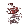
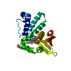
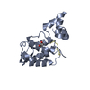
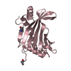
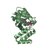
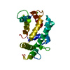
- PDB-7mtt: Crystal structure of colibactin self-resistance protein ClbS in c... -
+ Open data

- Basic information
| Entry | Database: PDB / ID: 7mtt | ||||||
|---|---|---|---|---|---|---|---|
| Title | Crystal structure of colibactin self-resistance protein ClbS in complex with two molecules of CHES | ||||||
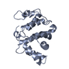
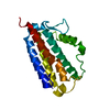
 Components Components | Colibactin self-protection protein ClbS | ||||||
 Keywords Keywords | DNA BINDING PROTEIN / hydrolase / DNA repair | ||||||
| Function / homology | Protein of unknown function DUF1706 / Protein of unknown function (DUF1706) / DinB/YfiT-like putative metalloenzymes / Colibactin self-protection protein ClbS Function and homology information Function and homology information | ||||||
| Biological species |  | ||||||
| Method |  X-RAY DIFFRACTION / SYNCHROTRON / MOLECULAR REPLACEMENT / Resolution: 1.85 Å X-RAY DIFFRACTION / SYNCHROTRON / MOLECULAR REPLACEMENT / Resolution: 1.85 Å | ||||||
 Authors Authors | Tripathi, P. / Bruner, S.D. | ||||||
 Citation Citation | Journal: Biochemistry / Year: 2021 Title: Structural Basis for the Interactions of the Colibactin Resistance Gene Product ClbS with DNA. Authors: Tripathi, P. / Bruner, S.D. #1: Journal: Journal of American Chemical Society / Year: 2017Title: ClbS Is a Cyclopropane Hydrolase That Confers Colibactin Resistance Authors: Tripathi, P. / Bruner, S.D. | ||||||
| History |
|
- Structure visualization
| Structure viewer | Molecule: MolmilJmol/JSmol |
|---|
- Downloads & links
-Download
| PDBx/mmCIF format | 7mtt.cif.gz | 51.8 KB | Display | PDBx/mmCIF format |
|---|---|---|---|---|
| PDB format | pdb7mtt.ent.gz | 34.2 KB | Display | PDB format |
| PDBx/mmJSON format | 7mtt.json.gz | Tree view | PDBx/mmJSON format | |
| Others |  Other downloads Other downloads |
-Validation report
| Arichive directory | https://data.pdbj.org/pub/pdb/validation_reports/mt/7mttftp://data.pdbj.org/pub/pdb/validation_reports/mt/7mtt | HTTPS FTP |
|---|
-Related structure data
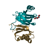
| Related structure data | 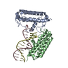 7mtlC  6anrS S: Starting model for refinement C: citing same article ( |
|---|---|
| Similar structure data |

-Links
 PDBj
PDBj- Assembly
| Deposited unit | 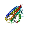
| ||||||||
|---|---|---|---|---|---|---|---|---|---|
| 1 |
| ||||||||
| Unit cell |
|
-Components
| #1: Protein | Mass: 20747.664 Da / Num. of mol.: 1 Source method: isolated from a genetically manipulated source Source: (gene. exp.) Gene: clbS, APU18_08100, AWF59_018730, AZZ83_004503, CT146_21495, CUB99_02165, D3C88_24730, DNR41_00045, DNX30_29170, DS966_21615, DU333_02935, DW236_02620, ELT23_23590, ELT33_24270, EPS70_02685, ...Gene: clbS, APU18_08100, AWF59_018730, AZZ83_004503, CT146_21495, CUB99_02165, D3C88_24730, DNR41_00045, DNX30_29170, DS966_21615, DU333_02935, DW236_02620, ELT23_23590, ELT33_24270, EPS70_02685, EPS94_00180, EWK56_23755, FPI65_12320, FQU83_11985, GFU47_04510, GP945_05010, GP946_20500, HHG54_004716, HHJ44_00090, HJL93_000008, HJM41_001166, HJO44_004707, HJS53_002303, HmCmsJML146_00176, HNX34_25610, HV055_09710, HV098_09980, HV348_09415, NCTC9075_02752, NCTC9434_01964 Production host: | ||||
|---|---|---|---|---|---|
| #2: Chemical |   Mass: 207.290 Da / Num. of mol.: 2 / Source method: obtained synthetically / Formula: C8H17NO3S / Feature type: SUBJECT OF INVESTIGATION / Comment: pH buffer*YM Mass: 207.290 Da / Num. of mol.: 2 / Source method: obtained synthetically / Formula: C8H17NO3S / Feature type: SUBJECT OF INVESTIGATION / Comment: pH buffer*YM#3: Water | ChemComp-HOH / |  Mass: 18.015 Da / Num. of mol.: 50 / Source method: isolated from a natural source / Formula: H2O Mass: 18.015 Da / Num. of mol.: 50 / Source method: isolated from a natural source / Formula: H2OHas ligand of interest | Y | |
-Experimental details
-Experiment
| Experiment | Method: X-RAY DIFFRACTION / Number of used crystals: 1 |
|---|
- Sample preparation
| Crystal | Density Matthews: 2.57 Å3/Da / Density % sol: 52.13 % |
|---|---|
| Crystal grow | Temperature: 277 K / Method: vapor diffusion, sitting drop / Details: 0.1 M CHES, pH 9.5, and 20% (w/v) PEG 8000 |
-Data collection
| Diffraction | Mean temperature: 100 K / Serial crystal experiment: N |
|---|---|
| Diffraction source | Source: SYNCHROTRON / Site: APS  / Beamline: 21-ID-G / Wavelength: 1.033 Å / Beamline: 21-ID-G / Wavelength: 1.033 Å |
| Detector | Type: MARMOSAIC 300 mm CCD / Detector: CCD / Date: Dec 5, 2019 |
| Radiation | Protocol: SINGLE WAVELENGTH / Monochromatic (M) / Laue (L): M / Scattering type: x-ray |
| Radiation wavelength | Wavelength: 1.033 Å / Relative weight: 1 |
| Reflection | Resolution: 1.85→44.62 Å / Num. obs: 17844 / % possible obs: 99.58 % / Redundancy: 6.4 % / CC1/2: 0.999 / Net I/σ(I): 19.4 |
| Reflection shell | Resolution: 1.85→1.915 Å / Num. unique obs: 1713 / CC1/2: 0.961 |
- Processing
| Software |
| ||||||||||||||||||||||||
|---|---|---|---|---|---|---|---|---|---|---|---|---|---|---|---|---|---|---|---|---|---|---|---|---|---|
| Refinement | Method to determine structure: MOLECULAR REPLACEMENT Starting model: 6ANR Resolution: 1.85→44.62 Å / Cor.coef. Fo:Fc: 0.96 / Cor.coef. Fo:Fc free: 0.941 / SU B: 3.265 / SU ML: 0.096 / Cross valid method: THROUGHOUT / σ(F): 0 / ESU R: 0.131 / ESU R Free: 0.129 / Stereochemistry target values: MAXIMUM LIKELIHOOD Details: HYDROGENS HAVE BEEN ADDED IN THE RIDING POSITIONS U VALUES : REFINED INDIVIDUALLY
| ||||||||||||||||||||||||
| Solvent computation | Ion probe radii: 0.8 Å / Shrinkage radii: 0.8 Å / VDW probe radii: 1.2 Å / Solvent model: MASK | ||||||||||||||||||||||||
| Displacement parameters | Biso max: 93.59 Å2 / Biso mean: 36.132 Å2 / Biso min: 19.37 Å2
| ||||||||||||||||||||||||
| Refinement step | Cycle: final / Resolution: 1.85→44.62 Å
| ||||||||||||||||||||||||
| LS refinement shell | Resolution: 1.85→1.897 Å / Rfactor Rfree error: 0
|
