 Movie
Movie Controller

[English] 日本語
 Yorodumi
- PDB-7mo8: Cryo-EM structure of 1:1 c-MET I/HGF I complex after focused 3D r... -
+ Open data

- Basic information
| Entry | Database: PDB / ID: 7mo8 | ||||||
|---|---|---|---|---|---|---|---|
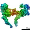
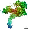
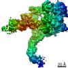
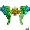
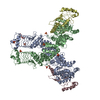
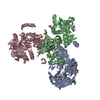
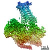
| Title | Cryo-EM structure of 1:1 c-MET I/HGF I complex after focused 3D refinement of holo-complex | ||||||
 Components Components |
| ||||||
 Keywords Keywords | SIGNALING PROTEIN / c-MET / HGF / receptor tyrosine kinase | ||||||
| Function / homology |  Function and homology information Function and homology informationregulation of branching involved in salivary gland morphogenesis by mesenchymal-epithelial signaling / hepatocyte growth factor receptor activity / Drug-mediated inhibition of MET activation / MET activates STAT3 / negative regulation of hydrogen peroxide-mediated programmed cell death / endothelial cell morphogenesis / MET Receptor Activation / MET interacts with TNS proteins / pancreas development / semaphorin receptor activity ...regulation of branching involved in salivary gland morphogenesis by mesenchymal-epithelial signaling / hepatocyte growth factor receptor activity / Drug-mediated inhibition of MET activation / MET activates STAT3 / negative regulation of hydrogen peroxide-mediated programmed cell death / endothelial cell morphogenesis / MET Receptor Activation / MET interacts with TNS proteins / pancreas development / semaphorin receptor activity / MET receptor recycling / cellular response to hepatocyte growth factor stimulus / MET activates PTPN11 / hepatocyte growth factor receptor signaling pathway / MET activates RAP1 and RAC1 / Sema4D mediated inhibition of cell attachment and migration / positive regulation of endothelial cell chemotaxis / MET activates PI3K/AKT signaling / MET activates PTK2 signaling / branching morphogenesis of an epithelial tube / positive regulation of DNA biosynthetic process / negative regulation of release of cytochrome c from mitochondria / positive chemotaxis / chemoattractant activity / positive regulation of osteoblast differentiation / semaphorin-plexin signaling pathway / epithelial to mesenchymal transition / Regulation of MITF-M-dependent genes involved in cell cycle and proliferation / MET activates RAS signaling / MECP2 regulates neuronal receptors and channels / platelet alpha granule lumen / Interleukin-7 signaling / cell surface receptor protein tyrosine kinase signaling pathway / liver development / basal plasma membrane / negative regulation of autophagy / excitatory postsynaptic potential / molecular function activator activity / InlB-mediated entry of Listeria monocytogenes into host cell / growth factor activity / cell chemotaxis / receptor protein-tyrosine kinase / Negative regulation of MET activity / Constitutive Signaling by Aberrant PI3K in Cancer / neuron differentiation / Platelet degranulation / PIP3 activates AKT signaling / mitotic cell cycle / PI5P, PP2A and IER3 Regulate PI3K/AKT Signaling / RAF/MAP kinase cascade / protein tyrosine kinase activity / Interleukin-4 and Interleukin-13 signaling / protein phosphatase binding / positive regulation of MAPK cascade / positive regulation of phosphatidylinositol 3-kinase/protein kinase B signal transduction / signaling receptor complex / cell surface receptor signaling pathway / postsynapse / positive regulation of cell migration / signaling receptor binding / negative regulation of apoptotic process / cell surface / positive regulation of transcription by RNA polymerase II / : / extracellular region / ATP binding / membrane / identical protein binding / plasma membrane Similarity search - Function | ||||||
| Biological species |  Homo sapiens (human) Homo sapiens (human) | ||||||
| Method | ELECTRON MICROSCOPY / single particle reconstruction / cryo EM / Resolution: 4.5 Å | ||||||
 Authors Authors | Uchikawa, E. / Chen, Z.M. / Xiao, G.Y. / Zhang, X.W. / Bai, X.C. | ||||||
 Citation Citation | Journal: Nat Commun / Year: 2021 Title: Structural basis of the activation of c-MET receptor. Authors: Emiko Uchikawa / Zhiming Chen / Guan-Yu Xiao / Xuewu Zhang / Xiao-Chen Bai /   Abstract: The c-MET receptor is a receptor tyrosine kinase (RTK) that plays essential roles in normal cell development and motility. Aberrant activation of c-MET can lead to both tumors growth and metastatic ...The c-MET receptor is a receptor tyrosine kinase (RTK) that plays essential roles in normal cell development and motility. Aberrant activation of c-MET can lead to both tumors growth and metastatic progression of cancer cells. C-MET can be activated by either hepatocyte growth factor (HGF), or its natural isoform NK1. Here, we report the cryo-EM structures of c-MET/HGF and c-MET/NK1 complexes in the active state. The c-MET/HGF complex structure reveals that, by utilizing two distinct interfaces, one HGF molecule is sufficient to induce a specific dimerization mode of c-MET for receptor activation. The binding of heparin as well as a second HGF to the 2:1 c-MET:HGF complex further stabilize this active conformation. Distinct to HGF, NK1 forms a stable dimer, and bridges two c-METs in a symmetrical manner for activation. Collectively, our studies provide structural insights into the activation mechanisms of c-MET, and reveal how two isoforms of the same ligand use dramatically different mechanisms to activate the receptor. | ||||||
| History |
|
- Structure visualization
| Movie |
Movie viewer |
|---|---|
| Structure viewer | Molecule: MolmilJmol/JSmol |
- Downloads & links
-Download
| PDBx/mmCIF format | 7mo8.cif.gz | 256.2 KB | Display | PDBx/mmCIF format |
|---|---|---|---|---|
| PDB format | pdb7mo8.ent.gz | 188.8 KB | Display | PDB format |
| PDBx/mmJSON format | 7mo8.json.gz | Tree view | PDBx/mmJSON format | |
| Others |  Other downloads Other downloads |
-Validation report
| Arichive directory | https://data.pdbj.org/pub/pdb/validation_reports/mo/7mo8ftp://data.pdbj.org/pub/pdb/validation_reports/mo/7mo8 | HTTPS FTP |
|---|
-Related structure data
| Related structure data | 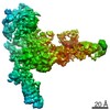 23920MC  7mo7C  7mo9C  7moaC 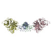 7mobC M: map data used to model this data C: citing same article ( |
|---|---|
| Similar structure data |


-Links
 PDBj
PDBj


- Assembly
| Deposited unit | 
|
|---|---|
| 1 |
|
-Components
| #1: Protein | Mass: 83249.828 Da / Num. of mol.: 1 Source method: isolated from a genetically manipulated source Source: (gene. exp.) Homo sapiens (human) / Gene: HGF, HPTA / Production host: Homo sapiens (human) / References: UniProt: P14210 |
|---|---|
| #2: Protein | Mass: 155720.625 Da / Num. of mol.: 1 Source method: isolated from a genetically manipulated source Source: (gene. exp.) Homo sapiens (human) / Gene: MET / Production host: Homo sapiens (human)References: UniProt: P08581, receptor protein-tyrosine kinase |
| #3: Polysaccharide | 2-O-sulfo-alpha-L-idopyranuronic acid-(1-4)-2-deoxy-6-O-sulfo-2-(sulfoamino)-alpha-D-glucopyranose- ...2-O-sulfo-alpha-L-idopyranuronic acid-(1-4)-2-deoxy-6-O-sulfo-2-(sulfoamino)-alpha-D-glucopyranose-(1-4)-2-O-sulfo-alpha-L-idopyranuronic acid-(1-4)-2-deoxy-6-O-sulfo-2-(sulfoamino)-alpha-D-glucopyranose-(1-4)-2-O-sulfo-alpha-L-idopyranuronic acid-(1-4)-2-deoxy-6-O-sulfo-2-(sulfoamino)-alpha-D-glucopyranose |
| Has ligand of interest | Y |
| Has protein modification | Y |
-Experimental details
-Experiment
| Experiment | Method: ELECTRON MICROSCOPY |
|---|---|
| EM experiment | Aggregation state: PARTICLE / 3D reconstruction method: single particle reconstruction |
- Sample preparation
| Component | Name: 1:1 c-MET I/HGF I complex / Type: COMPLEX / Entity ID: #1-#2 / Source: RECOMBINANT |
|---|---|
| Source (natural) | Organism: Homo sapiens (human) |
| Source (recombinant) | Organism: Homo sapiens (human) |
| Buffer solution | pH: 7.5 |
| Specimen | Embedding applied: NO / Shadowing applied: NO / Staining applied: NO / Vitrification applied: YES |
| Vitrification | Cryogen name: ETHANE |
- Electron microscopy imaging
| Experimental equipment |  Model: Titan Krios / Image courtesy: FEI Company |
|---|---|
| Microscopy | Model: FEI TITAN KRIOS |
| Electron gun | Electron source:  FIELD EMISSION GUN / Accelerating voltage: 300 kV / Illumination mode: FLOOD BEAM FIELD EMISSION GUN / Accelerating voltage: 300 kV / Illumination mode: FLOOD BEAM |
| Electron lens | Mode: BRIGHT FIELD / Alignment procedure: COMA FREE |
| Specimen holder | Cryogen: NITROGEN / Specimen holder model: FEI TITAN KRIOS AUTOGRID HOLDER |
| Image recording | Electron dose: 60 e/Å2 / Film or detector model: GATAN K3 BIOQUANTUM (6k x 4k) |
- Processing
| Software |
| ||||||||||||||||||
|---|---|---|---|---|---|---|---|---|---|---|---|---|---|---|---|---|---|---|---|
| EM software |
| ||||||||||||||||||
| CTF correction | Type: PHASE FLIPPING AND AMPLITUDE CORRECTION | ||||||||||||||||||
| 3D reconstruction | Resolution: 4.5 Å / Resolution method: FSC 0.143 CUT-OFF / Num. of particles: 25787 / Symmetry type: POINT | ||||||||||||||||||
| Refinement | Cross valid method: NONE |
