 Movie
Movie Controller

[English] 日本語
 Yorodumi
- EMDB-23920: Cryo-EM structure of 1:1 c-MET I/HGF I complex after focused 3D r... -
+ Open data

- Basic information
| Entry | Database: EMDB / ID: EMD-23920 | |||||||||
|---|---|---|---|---|---|---|---|---|---|---|
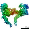
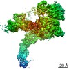
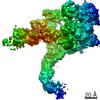
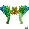
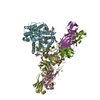
| Title | Cryo-EM structure of 1:1 c-MET I/HGF I complex after focused 3D refinement of holo-complex | |||||||||
 Map data Map data | Cryo-EM map of 1:1 c-MET I/HGF I complex after focused 3D refinement of holo-complex | |||||||||
 Sample Sample |
| |||||||||
 Keywords Keywords | c-MET / HGF / receptor tyrosine kinase / SIGNALING PROTEIN | |||||||||
| Function / homology |  Function and homology information Function and homology informationregulation of branching involved in salivary gland morphogenesis by mesenchymal-epithelial signaling / hepatocyte growth factor receptor activity / Drug-mediated inhibition of MET activation / MET activates STAT3 / negative regulation of hydrogen peroxide-mediated programmed cell death / endothelial cell morphogenesis / MET Receptor Activation / MET interacts with TNS proteins / pancreas development / semaphorin receptor activity ...regulation of branching involved in salivary gland morphogenesis by mesenchymal-epithelial signaling / hepatocyte growth factor receptor activity / Drug-mediated inhibition of MET activation / MET activates STAT3 / negative regulation of hydrogen peroxide-mediated programmed cell death / endothelial cell morphogenesis / MET Receptor Activation / MET interacts with TNS proteins / pancreas development / semaphorin receptor activity / MET receptor recycling / cellular response to hepatocyte growth factor stimulus / MET activates PTPN11 / hepatocyte growth factor receptor signaling pathway / MET activates RAP1 and RAC1 / Sema4D mediated inhibition of cell attachment and migration / positive regulation of endothelial cell chemotaxis / MET activates PI3K/AKT signaling / MET activates PTK2 signaling / branching morphogenesis of an epithelial tube / positive regulation of DNA biosynthetic process / negative regulation of release of cytochrome c from mitochondria / positive chemotaxis / chemoattractant activity / positive regulation of osteoblast differentiation / semaphorin-plexin signaling pathway / epithelial to mesenchymal transition / Regulation of MITF-M-dependent genes involved in cell cycle and proliferation / MET activates RAS signaling / MECP2 regulates neuronal receptors and channels / platelet alpha granule lumen / Interleukin-7 signaling / cell surface receptor protein tyrosine kinase signaling pathway / liver development / basal plasma membrane / negative regulation of autophagy / excitatory postsynaptic potential / molecular function activator activity / InlB-mediated entry of Listeria monocytogenes into host cell / growth factor activity / cell chemotaxis / receptor protein-tyrosine kinase / Negative regulation of MET activity / Constitutive Signaling by Aberrant PI3K in Cancer / neuron differentiation / Platelet degranulation / PIP3 activates AKT signaling / mitotic cell cycle / PI5P, PP2A and IER3 Regulate PI3K/AKT Signaling / RAF/MAP kinase cascade / protein tyrosine kinase activity / Interleukin-4 and Interleukin-13 signaling / protein phosphatase binding / positive regulation of MAPK cascade / positive regulation of phosphatidylinositol 3-kinase/protein kinase B signal transduction / signaling receptor complex / cell surface receptor signaling pathway / postsynapse / positive regulation of cell migration / signaling receptor binding / negative regulation of apoptotic process / cell surface / positive regulation of transcription by RNA polymerase II / : / extracellular region / ATP binding / membrane / identical protein binding / plasma membrane Similarity search - Function | |||||||||
| Biological species |  Homo sapiens (human) Homo sapiens (human) | |||||||||
| Method | single particle reconstruction / cryo EM / Resolution: 4.5 Å | |||||||||
 Authors Authors | Uchikawa E / Chen ZM | |||||||||
 Citation Citation | Journal: Nat Commun / Year: 2021 Title: Structural basis of the activation of c-MET receptor. Authors: Emiko Uchikawa / Zhiming Chen / Guan-Yu Xiao / Xuewu Zhang / Xiao-Chen Bai /   Abstract: The c-MET receptor is a receptor tyrosine kinase (RTK) that plays essential roles in normal cell development and motility. Aberrant activation of c-MET can lead to both tumors growth and metastatic ...The c-MET receptor is a receptor tyrosine kinase (RTK) that plays essential roles in normal cell development and motility. Aberrant activation of c-MET can lead to both tumors growth and metastatic progression of cancer cells. C-MET can be activated by either hepatocyte growth factor (HGF), or its natural isoform NK1. Here, we report the cryo-EM structures of c-MET/HGF and c-MET/NK1 complexes in the active state. The c-MET/HGF complex structure reveals that, by utilizing two distinct interfaces, one HGF molecule is sufficient to induce a specific dimerization mode of c-MET for receptor activation. The binding of heparin as well as a second HGF to the 2:1 c-MET:HGF complex further stabilize this active conformation. Distinct to HGF, NK1 forms a stable dimer, and bridges two c-METs in a symmetrical manner for activation. Collectively, our studies provide structural insights into the activation mechanisms of c-MET, and reveal how two isoforms of the same ligand use dramatically different mechanisms to activate the receptor. | |||||||||
| History |
|
- Structure visualization
| Movie |
Movie viewer |
|---|---|
| Structure viewer | EM map: SurfViewMolmilJmol/JSmol |
| Supplemental images |

- Downloads & links
-EMDB archive
| Map data | emd_23920.map.gz | 70.2 MB | EMDB map data format | |
|---|---|---|---|---|
| Header (meta data) | emd-23920-v30.xmlemd-23920.xml | 12.7 KB 12.7 KB | Display Display | EMDB header |
| Images | 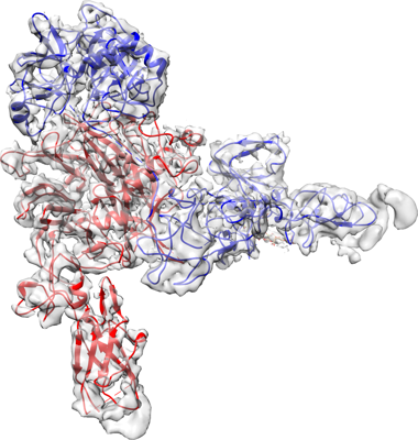 emd_23920.png emd_23920.png | 161.1 KB | ||
| Filedesc metadata | emd-23920.cif.gz | 6.5 KB | ||
| Archive directory |  http://ftp.pdbj.org/pub/emdb/structures/EMD-23920ftp://ftp.pdbj.org/pub/emdb/structures/EMD-23920 http://ftp.pdbj.org/pub/emdb/structures/EMD-23920ftp://ftp.pdbj.org/pub/emdb/structures/EMD-23920 | HTTPS FTP |
-Related structure data
| Related structure data |  7mo8MC  7mo7C  7mo9C  7moaC  7mobC M: atomic model generated by this map C: citing same article ( |
|---|---|
| Similar structure data |


-Links
| EMDB pages | EMDB (EBI/PDBe) / EMDataResource |
|---|---|
| Related items in Molecule of the Month |


-Map
| File | Download / File: emd_23920.map.gz / Format: CCP4 / Size: 75.1 MB / Type: IMAGE STORED AS FLOATING POINT NUMBER (4 BYTES) | ||||||||||||||||||||||||||||||||||||||||||||||||||||||||||||||||||||
|---|---|---|---|---|---|---|---|---|---|---|---|---|---|---|---|---|---|---|---|---|---|---|---|---|---|---|---|---|---|---|---|---|---|---|---|---|---|---|---|---|---|---|---|---|---|---|---|---|---|---|---|---|---|---|---|---|---|---|---|---|---|---|---|---|---|---|---|---|---|
| Annotation | Cryo-EM map of 1:1 c-MET I/HGF I complex after focused 3D refinement of holo-complex | ||||||||||||||||||||||||||||||||||||||||||||||||||||||||||||||||||||
| Projections & slices | Image control
Images are generated by Spider. | ||||||||||||||||||||||||||||||||||||||||||||||||||||||||||||||||||||
| Voxel size | X=Y=Z: 1.08 Å | ||||||||||||||||||||||||||||||||||||||||||||||||||||||||||||||||||||
| Density |
| ||||||||||||||||||||||||||||||||||||||||||||||||||||||||||||||||||||
| Symmetry | Space group: 1 | ||||||||||||||||||||||||||||||||||||||||||||||||||||||||||||||||||||
| Details | EMDB XML:
CCP4 map header:
| ||||||||||||||||||||||||||||||||||||||||||||||||||||||||||||||||||||
 Z (Sec.)
 Y (Row.)
 X (Col.)


-Supplemental data
- Sample components
-Entire : 1:1 c-MET I/HGF I complex
| Entire | Name: 1:1 c-MET I/HGF I complex |
|---|---|
| Components |
|
-Supramolecule #1: 1:1 c-MET I/HGF I complex
| Supramolecule | Name: 1:1 c-MET I/HGF I complex / type: complex / ID: 1 / Parent: 0 / Macromolecule list: all |
|---|---|
| Source (natural) | Organism: Homo sapiens (human) |
-Macromolecule #1: Hepatocyte growth factor
| Macromolecule | Name: Hepatocyte growth factor / type: protein_or_peptide / ID: 1 / Number of copies: 1 / Enantiomer: LEVO |
|---|---|
| Source (natural) | Organism: Homo sapiens (human) |
| Molecular weight | Theoretical: 83.249828 KDa |
| Recombinant expression | Organism: Homo sapiens (human) |
| Sequence | String: MWVTKLLPAL LLQHVLLHLL LLPIAIPYAE GQRKRRNTIH EFKKSAKTTL IKIDPALKIK TKKVNTADQC ANRCTRNKGL PFTCKAFVF DKARKQCLWF PFNSMSSGVK KEFGHEFDLY ENKDYIRNCI IGKGRSYKGT VSITKSGIKC QPWSSMIPHE H SFLPSSYR ...String: MWVTKLLPAL LLQHVLLHLL LLPIAIPYAE GQRKRRNTIH EFKKSAKTTL IKIDPALKIK TKKVNTADQC ANRCTRNKGL PFTCKAFVF DKARKQCLWF PFNSMSSGVK KEFGHEFDLY ENKDYIRNCI IGKGRSYKGT VSITKSGIKC QPWSSMIPHE H SFLPSSYR GKDLQENYCR NPRGEEGGPW CFTSNPEVRY EVCDIPQCSE VECMTCNGES YRGLMDHTES GKICQRWDHQ TP HRHKFLP ERYPDKGFDD NYCRNPDGQP RPWCYTLDPH TRWEYCAIKT CADNTMNDTD VPLETTECIQ GQGEGYRGTV NTI WNGIPC QRWDSQYPHE HDMTPENFKC KDLRENYCRN PDGSESPWCF TTDPNIRVGY CSQIPNCDMS HGQDCYRGNG KNYM GNLSQ TRSGLTCSMW DKNMEDLHRH IFWEPDASKL NENYCRNPDD DAHGPWCYTG NPLIPWDYCP ISRCEGDTTP TIVNL DHPV ISCAKTKQLR VVNGIPTRTN IGWMVSLRYR NKHICGGSLI KESWVLTARQ CFPSRDLKDY EAWLGIHDVH GRGDEK CKQ VLNVSQLVYG PEGSDLVLMK LARPAVLDDF VSTIDLPNYG CTIPEKTSCS VYGWGYTGLI NYDGLLRVAH LYIMGNE KC SQHHRGKVTL NESEICAGAE KIGSGPCEGD YGGPLVCEQH KMRMVLGVIV PGRGCAIPNR PGIFVRVAYY AKWIHKII L TYKVPQS UniProtKB: Hepatocyte growth factor |
-Macromolecule #2: Hepatocyte growth factor receptor
| Macromolecule | Name: Hepatocyte growth factor receptor / type: protein_or_peptide / ID: 2 / Number of copies: 1 / Enantiomer: LEVO / EC number: receptor protein-tyrosine kinase |
|---|---|
| Source (natural) | Organism: Homo sapiens (human) |
| Molecular weight | Theoretical: 155.720625 KDa |
| Recombinant expression | Organism: Homo sapiens (human) |
| Sequence | String: MKAPAVLAPG ILVLLFTLVQ RSNGECKEAL AKSEMNVNMK YQLPNFTAET PIQNVILHEH HIFLGATNYI YVLNEEDLQK VAEYKTGPV LEHPDCFPCQ DCSSKANLSG GVWKDNINMA LVVDTYYDDQ LISCGSVNRG TCQRHVFPHN HTADIQSEVH C IFSPQIEE ...String: MKAPAVLAPG ILVLLFTLVQ RSNGECKEAL AKSEMNVNMK YQLPNFTAET PIQNVILHEH HIFLGATNYI YVLNEEDLQK VAEYKTGPV LEHPDCFPCQ DCSSKANLSG GVWKDNINMA LVVDTYYDDQ LISCGSVNRG TCQRHVFPHN HTADIQSEVH C IFSPQIEE PSQCPDCVVS ALGAKVLSSV KDRFINFFVG NTINSSYFPD HPLHSISVRR LKETKDGFMF LTDQSYIDVL PE FRDSYPI KYVHAFESNN FIYFLTVQRE TLDAQTFHTR IIRFCSINSG LHSYMEMPLE CILTEKRKKR STKKEVFNIL QAA YVSKPG AQLARQIGAS LNDDILFGVF AQSKPDSAEP MDRSAMCAFP IKYVNDFFNK IVNKNNVRCL QHFYGPNHEH CFNR TLLRN SSGCEARRDE YRTEFTTALQ RVDLFMGQFS EVLLTSISTF IKGDLTIANL GTSEGRFMQV VVSRSGPSTP HVNFL LDSH PVSPEVIVEH TLNQNGYTLV ITGKKITKIP LNGLGCRHFQ SCSQCLSAPP FVQCGWCHDK CVRSEECLSG TWTQQI CLP AIYKVFPNSA PLEGGTRLTI CGWDFGFRRN NKFDLKKTRV LLGNESCTLT LSESTMNTLK CTVGPAMNKH FNMSIII SN GHGTTQYSTF SYVDPVITSI SPKYGPMAGG TLLTLTGNYL NSGNSRHISI GGKTCTLKSV SNSILECYTP AQTISTEF A VKLKIDLANR ETSIFSYRED PIVYEIHPTK SFISGGSTIT GVGKNLNSVS VPRMVINVHE AGRNFTVACQ HRSNSEIIC CTTPSLQQLN LQLPLKTKAF FMLDGILSKY FDLIYVHNPV FKPFEKPVMI SMGNENVLEI KGNDIDPEAV KGEVLKVGNK SCENIHLHS EAVLCTVPND LLKLNSELNI EWKQAISSTV LGKVIVQPDQ NFTGLIAGVV SISTALLLLL GFFLWLKKRK Q IKDLGSEL VRYDARVHTP HLDRLVSARS VSPTTEMVSN ESVDYRATFP EDQFPNSSQN GSCRQVQYPL TDMSPILTSG DS DISSPLL QNTVHIDLSA LNPELVQAVQ HVVIGPSSLI VHFNEVIGRG HFGCVYHGTL LDNDGKKIHC AVKSLNRITD IGE VSQFLT EGIIMKDFSH PNVLSLLGIC LRSEGSPLVV LPYMKHGDLR NFIRNETHNP TVKDLIGFGL QVAKGMKYLA SKKF VHRDL AARNCMLDEK FTVKVADFGL ARDMYDKEYY SVHNKTGAKL PVKWMALESL QTQKFTTKSD VWSFGVLLWE LMTRG APPY PDVNTFDITV YLLQGRRLLQ PEYCPDPLYE VMLKCWHPKA EMRPSFSELV SRISAIFSTF IGEHYVHVNA TYVNVK CVA PYPSLLSSED NADDEVDTRP ASFWETS UniProtKB: Hepatocyte growth factor receptor |
-Experimental details
-Structure determination
| Method | cryo EM |
|---|---|
 Processing Processing | single particle reconstruction |
| Aggregation state | particle |
-Sample preparation
| Buffer | pH: 7.5 |
|---|---|
| Vitrification | Cryogen name: ETHANE |
- Electron microscopy
| Microscope | FEI TITAN KRIOS |
|---|---|
| Image recording | Film or detector model: GATAN K3 BIOQUANTUM (6k x 4k) / Average electron dose: 60.0 e/Å2 |
| Electron beam | Acceleration voltage: 300 kV / Electron source:  FIELD EMISSION GUN FIELD EMISSION GUN |
| Electron optics | Illumination mode: FLOOD BEAM / Imaging mode: BRIGHT FIELD |
| Sample stage | Specimen holder model: FEI TITAN KRIOS AUTOGRID HOLDER / Cooling holder cryogen: NITROGEN |
| Experimental equipment |  Model: Titan Krios / Image courtesy: FEI Company |
