 Movie
Movie Controller


+ Open data

- Basic information
| Entry | Database: EMDB / ID: EMD-22328 | |||||||||
|---|---|---|---|---|---|---|---|---|---|---|
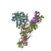
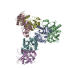
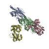
| Title | IgA1 Protease in complex with neutralizing mAb | |||||||||
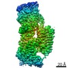
 Map data Map data | IgA1 Protease with mAb | |||||||||
 Sample Sample |
| |||||||||
 Keywords Keywords | IgA1 / Protease / neutralization / IMMUNE SYSTEM | |||||||||
| Function / homology |  Function and homology information Function and homology informationIgA-specific metalloendopeptidase / metalloendopeptidase activity / proteolysis / extracellular region / zinc ion binding / membrane Similarity search - Function | |||||||||
| Biological species |   Streptococcus pneumoniae (bacteria) / Streptococcus pneumoniae (bacteria) /  | |||||||||
| Method | single particle reconstruction / cryo EM / Resolution: 4.8 Å | |||||||||
 Authors Authors | Eisenmesser EZ / Zheng H | |||||||||
| Funding support |  United States, 1 items United States, 1 items
| |||||||||
 Citation Citation | Journal: Nat Commun / Year: 2020 Title: Mechanism and inhibition of Streptococcus pneumoniae IgA1 protease. Authors: Zhiming Wang / Jeremy Rahkola / Jasmina S Redzic / Ying-Chih Chi / Norman Tran / Todd Holyoak / Hongjin Zheng / Edward Janoff / Elan Eisenmesser /  Abstract: Opportunistic pathogens such as Streptococcus pneumoniae secrete a giant metalloprotease virulence factor responsible for cleaving host IgA1, yet the molecular mechanism has remained unknown since ...Opportunistic pathogens such as Streptococcus pneumoniae secrete a giant metalloprotease virulence factor responsible for cleaving host IgA1, yet the molecular mechanism has remained unknown since their discovery nearly 30 years ago despite the potential for developing vaccines that target these enzymes to block infection. Here we show through a series of cryo-electron microscopy single particle reconstructions how the Streptococcus pneumoniae IgA1 protease facilitates IgA1 substrate recognition and how this can be inhibited. Specifically, the Streptococcus pneumoniae IgA1 protease subscribes to an active-site-gated mechanism where a domain undergoes a 10.0 Å movement to facilitate cleavage. Monoclonal antibody binding inhibits this conformational change, providing a direct means to block infection at the host interface. These structural studies explain decades of biological and biochemical studies and provides a general strategy to block Streptococcus pneumoniae IgA1 protease activity to potentially prevent infection. | |||||||||
| History |
|
- Structure visualization
| Movie |
Movie viewer |
|---|---|
| Structure viewer | EM map: SurfViewMolmilJmol/JSmol |
| Supplemental images |
 UCSF Chimera
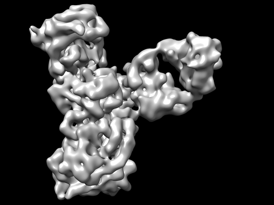
- Downloads & links
-EMDB archive
| Map data | emd_22328.map.gz | 122.3 MB | EMDB map data format | |
|---|---|---|---|---|
| Header (meta data) | emd-22328-v30.xmlemd-22328.xml | 18 KB 18 KB | Display Display | EMDB header |
| Images |  emd_22328.png emd_22328.png | 72.1 KB | ||
| Filedesc metadata | emd-22328.cif.gz | 6.9 KB | ||
| Archive directory |  http://ftp.pdbj.org/pub/emdb/structures/EMD-22328ftp://ftp.pdbj.org/pub/emdb/structures/EMD-22328 http://ftp.pdbj.org/pub/emdb/structures/EMD-22328ftp://ftp.pdbj.org/pub/emdb/structures/EMD-22328 | HTTPS FTP |
-Related structure data
| Related structure data |  7jgjMC  6xjaC 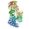 6xjbC M: atomic model generated by this map C: citing same article ( |
|---|---|
| Similar structure data |


-Links
| EMDB pages | EMDB (EBI/PDBe) / EMDataResource |
|---|
-Map
| File | Download / File: emd_22328.map.gz / Format: CCP4 / Size: 129.7 MB / Type: IMAGE STORED AS FLOATING POINT NUMBER (4 BYTES) | ||||||||||||||||||||||||||||||||||||||||||||||||||||||||||||||||||||
|---|---|---|---|---|---|---|---|---|---|---|---|---|---|---|---|---|---|---|---|---|---|---|---|---|---|---|---|---|---|---|---|---|---|---|---|---|---|---|---|---|---|---|---|---|---|---|---|---|---|---|---|---|---|---|---|---|---|---|---|---|---|---|---|---|---|---|---|---|---|
| Annotation | IgA1 Protease with mAb | ||||||||||||||||||||||||||||||||||||||||||||||||||||||||||||||||||||
| Projections & slices | Image control
Images are generated by Spider. | ||||||||||||||||||||||||||||||||||||||||||||||||||||||||||||||||||||
| Voxel size | X=Y=Z: 0.832 Å | ||||||||||||||||||||||||||||||||||||||||||||||||||||||||||||||||||||
| Density |
| ||||||||||||||||||||||||||||||||||||||||||||||||||||||||||||||||||||
| Symmetry | Space group: 1 | ||||||||||||||||||||||||||||||||||||||||||||||||||||||||||||||||||||
| Details | EMDB XML:
CCP4 map header:
| ||||||||||||||||||||||||||||||||||||||||||||||||||||||||||||||||||||
 Z (Sec.)
 Y (Row.)
 X (Col.)


-Supplemental data
- Sample components
-Entire : murine mAb in complex with Streptoccocus pneumoniae IgA1 Protease
| Entire | Name: murine mAb in complex with Streptoccocus pneumoniae IgA1 Protease |
|---|---|
| Components |
|
-Supramolecule #1: murine mAb in complex with Streptoccocus pneumoniae IgA1 Protease
| Supramolecule | Name: murine mAb in complex with Streptoccocus pneumoniae IgA1 Protease type: complex / ID: 1 / Parent: 0 / Macromolecule list: all |
|---|
-Supramolecule #2: Immunoglobulin A1 protease
| Supramolecule | Name: Immunoglobulin A1 protease / type: complex / ID: 2 / Parent: 1 / Macromolecule list: #1 |
|---|---|
| Source (natural) | Organism: Streptococcus pneumoniae (bacteria) |
-Supramolecule #3: mAb
| Supramolecule | Name: mAb / type: complex / ID: 3 / Parent: 1 / Macromolecule list: #2-#3 |
|---|---|
| Source (natural) | Organism: |
-Macromolecule #1: Immunoglobulin A1 protease
| Macromolecule | Name: Immunoglobulin A1 protease / type: protein_or_peptide / ID: 1 / Number of copies: 1 / Enantiomer: LEVO |
|---|---|
| Source (natural) | Organism: Streptococcus pneumoniae (bacteria) |
| Molecular weight | Theoretical: 145.095812 KDa |
| Recombinant expression | Organism: |
| Sequence | String: TEPEKKLELR NVSDIELYSQ TNGTYRQHVS LDGIPENTDT YFVKVKSSAF KDVYIPVASI TEEKRNGQSV YKITAKAEKL QQELENKYV DNFTFYLDKK AKEENTNFTS FSNLVKAINQ NPSGTYHLAA SLNANEVELG PDERSYIKDT FTGRLIGEKD G KNYAIYNL ...String: TEPEKKLELR NVSDIELYSQ TNGTYRQHVS LDGIPENTDT YFVKVKSSAF KDVYIPVASI TEEKRNGQSV YKITAKAEKL QQELENKYV DNFTFYLDKK AKEENTNFTS FSNLVKAINQ NPSGTYHLAA SLNANEVELG PDERSYIKDT FTGRLIGEKD G KNYAIYNL KKPLFENLSG ATVEKLSLKN VAISGKNDIG SLANEATNGT KIKQVHVDGV LAGERGVGGL LAKADQSSIA ES SFKGRIV NTYETTDAYN IGGLVGHLTG KNASIAKSKA TVTISSNTNR SDQTVGGLAG LVDQDAHIQN SYAEGDINNV KHF GKVAGV AGYLWDRTSG EEKHAGELTN VLSDVNVTNG NAITGYHYTG MKVANTFSSK ANRVFNVTLE KDEVVSKESF EERG TMLDA SQIVSKKAEI NPLTLPTVEP LSTSGKKDSD FSKIAHYQAN RALVYKNIEK LLPFYNKSTI VKYGNLVKEN SLLYQ KELL SAVMMKDDQV ITDIVSNKQT ANKLLLHYND HSSEKFDLKY QTDFANLAEY NLGNTGLLYT PNQFLYDRDS IVKEVL PEL QKLDYQSDAI RKTLGISPEV KLTELYLEDQ FSKTKQNLGD SLKKLLSADA GLASDNSVTR GYLVDKIKNN KEALLLG LT YLERWYNFNY GQVNVKDLVM YHPDFFGKGN TSPLDTLIEL GKSGFNNLLA KNNVDTYGIS LASQHGATDL FSTLEHYR K VFLPNTSNND WFKSETKAYI VEEKSTIEEV KTKQGLAGTK YSIGVYDRIT SATWKYRNMV LPLLTLPERS VFVISTMSS LGFGAYDRYR SSDHKAGKAL NDFVEENARE TAKRQRDHYD YWYRILDEQS REKLYRTILL YDAYKFGDDT TSGKATAEAK FDSSNPAMK NFFGPVGNKV VHNQHGAYAT GDGVYYMSYR MLDKDGAITY THEMTHDSDQ DIYLGGYGRR NGLGPEFFAK G LLQAPDQP SDATITINSI LKHSKSDSTE GSRLQVLDPT ERFQNAADLQ NYVHNMFDLI YMMEYLEGQS IVNKLSVYQK MA ALRKIEN KYVKDPADGN EVYATNVVKE LTEAEARNLN SFESLIDHNI LSAREYQSGD YERNGYYTIK LFAPIYSALS SEK GTPGDL MGRRIAYELL AAKGFKDGMV PYISNQYEED AKQQGQTINL YGKERGLVTD ELVLKKVFDG KYKTWAEFKT AMYQ ERVDQ FGNLKQVTFK DPTKPWPSYG TKTINNVDEL QALMDQAVLK DAEGPRWSNY DPEIDSAVHK LKRAIFKAYL DQTND FRSS IFENKK UniProtKB: Immunoglobulin A1 protease |
-Macromolecule #2: mAB Light Chain
| Macromolecule | Name: mAB Light Chain / type: protein_or_peptide / ID: 2 / Number of copies: 1 / Enantiomer: LEVO |
|---|---|
| Source (natural) | Organism: |
| Molecular weight | Theoretical: 23.270615 KDa |
| Recombinant expression | Organism:  Homo sapiens (human) Homo sapiens (human) |
| Sequence | String: EEVLTQSPAI MSASPGEKVT MTCSASSSVS YIHWYQQKSN TSPKLWIYAT SKLASGVPGR FSGSGSGNSY SLTISSMEAE DVATYYCFQ GSGYPFTFGS GTKLEIKRAD AAPTVSIFPP SSEQLTSGGA SVVCFLNNFY PKDINVKWKI DGSERQNGVL N SWTDQDSK ...String: EEVLTQSPAI MSASPGEKVT MTCSASSSVS YIHWYQQKSN TSPKLWIYAT SKLASGVPGR FSGSGSGNSY SLTISSMEAE DVATYYCFQ GSGYPFTFGS GTKLEIKRAD AAPTVSIFPP SSEQLTSGGA SVVCFLNNFY PKDINVKWKI DGSERQNGVL N SWTDQDSK DSTYSMSSTL TLTKDEYERH NSYTCEATHK TSTSPIVKSF NRNEC |
-Macromolecule #3: mAB Heavy Chain
| Macromolecule | Name: mAB Heavy Chain / type: protein_or_peptide / ID: 3 / Number of copies: 1 / Enantiomer: LEVO |
|---|---|
| Source (natural) | Organism: |
| Molecular weight | Theoretical: 23.212004 KDa |
| Recombinant expression | Organism: Homo sapiens (human) |
| Sequence | String: EVQLQQSGPE LEKPGASMKI SCKASGYSFT GYNMNWVKQS NGKSLEWIGS IDPYTGGSNY NQKFMGKATL TVDKSSSTAY MQLKSLTSE DSAVYYCATV VGRVYAMDYW GQGTSVTVSS AKTTPPSVYP LAPGSAAQTN SMVTLGCLVK GYFPEPVTVT W NSGSLSSG ...String: EVQLQQSGPE LEKPGASMKI SCKASGYSFT GYNMNWVKQS NGKSLEWIGS IDPYTGGSNY NQKFMGKATL TVDKSSSTAY MQLKSLTSE DSAVYYCATV VGRVYAMDYW GQGTSVTVSS AKTTPPSVYP LAPGSAAQTN SMVTLGCLVK GYFPEPVTVT W NSGSLSSG VHTFPAVLQS DLYTLSSSVT VPSSTWPSET VTCNVAHPAS STKVDKKIVP |
-Experimental details
-Structure determination
| Method | cryo EM |
|---|---|
 Processing Processing | single particle reconstruction |
| Aggregation state | particle |
-Sample preparation
| Concentration | 1 mg/mL |
|---|---|
| Buffer | pH: 7.4 / Component - Concentration: 20.0 millimolar / Component - Name: HEPES / Details: 20 mM HEPES, pH 7.4 150 mM NaCl |
| Vitrification | Cryogen name: ETHANE |
- Electron microscopy
| Microscope | FEI TITAN KRIOS |
|---|---|
| Image recording | Film or detector model: GATAN K2 BASE (4k x 4k) / Average electron dose: 30.0 e/Å2 |
| Electron beam | Acceleration voltage: 300 kV / Electron source: OTHER |
| Electron optics | Illumination mode: OTHER / Imaging mode: OTHER |
| Experimental equipment |  Model: Titan Krios / Image courtesy: FEI Company |
