 Movie
Movie Controller

[English] 日本語
 Yorodumi
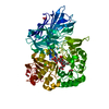
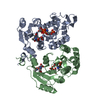
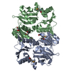
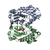
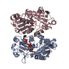
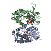
- PDB-5dmx: Crystal structure of D-alanine-D-alanine ligase from Acinetobacte... -
+ Open data

- Basic information
| Entry | Database: PDB / ID: 5dmx | ||||||
|---|---|---|---|---|---|---|---|
| Title | Crystal structure of D-alanine-D-alanine ligase from Acinetobacter baumannii, space group p212121 | ||||||
 Components Components | D-alanine--D-alanine ligase | ||||||
 Keywords Keywords | LIGASE / D-alanine-D-alanine ligase / Acinetobacter baumannii / drug target protein / apo structure | ||||||
| Function / homology |  Function and homology information Function and homology informationD-alanine-D-alanine ligase / D-alanine-D-alanine ligase activity / peptidoglycan biosynthetic process / cell wall organization / regulation of cell shape / ATP binding / metal ion binding / cytosol Similarity search - Function | ||||||
| Biological species |  Acinetobacter baumannii ACICU (bacteria) Acinetobacter baumannii ACICU (bacteria) | ||||||
| Method |  X-RAY DIFFRACTION / SYNCHROTRON / MOLECULAR REPLACEMENT / Resolution: 2.81 Å X-RAY DIFFRACTION / SYNCHROTRON / MOLECULAR REPLACEMENT / Resolution: 2.81 Å | ||||||
 Authors Authors | Huynh, K.H. / Hong, M.K. / Kang, L.W. | ||||||
 Citation Citation | Journal: J. Microbiol. / Year: 2015 Title: The crystal structure of the D-alanine-D-alanine ligase from Acinetobacter baumannii suggests a flexible conformational change in the central domain before nucleotide binding Authors: Huynh, K.H. / Hong, M.K. / Lee, C. / Tran, H.T. / Lee, S.H. / Ahn, Y.J. / Cha, S.S. / Kang, L.W. | ||||||
| History |
|
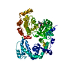
- Structure visualization
| Structure viewer | Molecule: MolmilJmol/JSmol |
|---|
- Downloads & links
-Download
| PDBx/mmCIF format | 5dmx.cif.gz | 285.3 KB | Display | PDBx/mmCIF format |
|---|---|---|---|---|
| PDB format | pdb5dmx.ent.gz | 231.1 KB | Display | PDB format |
| PDBx/mmJSON format | 5dmx.json.gz | Tree view | PDBx/mmJSON format | |
| Others |  Other downloads Other downloads |
-Validation report
| Arichive directory | https://data.pdbj.org/pub/pdb/validation_reports/dm/5dmxftp://data.pdbj.org/pub/pdb/validation_reports/dm/5dmx | HTTPS FTP |
|---|
-Related structure data
| Related structure data | 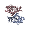 5d8dC 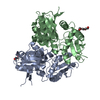 3v4zS S: Starting model for refinement C: citing same article ( |
|---|---|
| Similar structure data |


-Links
 PDBj
PDBj


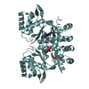
- Assembly
| Deposited unit | 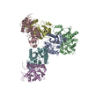
| ||||||||
|---|---|---|---|---|---|---|---|---|---|
| 1 | 
| ||||||||
| 2 | 
| ||||||||
| 3 | 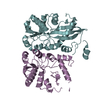
| ||||||||
| Unit cell |
|
-Components
| #1: Protein | Mass: 33392.746 Da / Num. of mol.: 6 Source method: isolated from a genetically manipulated source Source: (gene. exp.) Acinetobacter baumannii ACICU (bacteria)Strain: ACICU / Gene: ddl, ACICU_03532 / Production host: #2: Water | ChemComp-HOH / |  Mass: 18.015 Da / Num. of mol.: 69 / Source method: isolated from a natural source / Formula: H2O Mass: 18.015 Da / Num. of mol.: 69 / Source method: isolated from a natural source / Formula: H2O |
|---|
-Experimental details
-Experiment
| Experiment | Method: X-RAY DIFFRACTION / Number of used crystals: 1 |
|---|
- Sample preparation
| Crystal | Density Matthews: 2.94 Å3/Da / Density % sol: 58.15 % |
|---|---|
| Crystal grow | Temperature: 287 K / Method: vapor diffusion, sitting drop / pH: 6.5 Details: 0.06 M MgCl2, CaCl2, 0.1 M imidazole, MES-HCl, 30% EDO-P8K, 40%(v/v) ethylene glycol, 20%(w/v) polyethylene glycol 8000 |
-Data collection
| Diffraction | Mean temperature: 100 K |
|---|---|
| Diffraction source | Source: SYNCHROTRON / Site: PAL/PLS  / Beamline: 7A (6B, 6C1) / Wavelength: 0.979 Å / Beamline: 7A (6B, 6C1) / Wavelength: 0.979 Å |
| Detector | Type: ADSC QUANTUM 270 / Detector: CCD / Date: Nov 30, 2013 |
| Radiation | Protocol: SINGLE WAVELENGTH / Monochromatic (M) / Laue (L): M / Scattering type: x-ray |
| Radiation wavelength | Wavelength: 0.979 Å / Relative weight: 1 |
| Reflection | Resolution: 2.8→50 Å / Num. obs: 55982 / % possible obs: 96 % / Redundancy: 7.4 % / Net I/σ(I): 12.9 |
| Reflection shell | Resolution: 2.8→2.85 Å / Redundancy: 3.7 % / Rmerge(I) obs: 0.42 / Mean I/σ(I) obs: 1.6 / % possible all: 92 |
- Processing
| Software |
| |||||||||||||||||||||||||||||||||||||||||||||||||||||||||||||||||||||||||||
|---|---|---|---|---|---|---|---|---|---|---|---|---|---|---|---|---|---|---|---|---|---|---|---|---|---|---|---|---|---|---|---|---|---|---|---|---|---|---|---|---|---|---|---|---|---|---|---|---|---|---|---|---|---|---|---|---|---|---|---|---|---|---|---|---|---|---|---|---|---|---|---|---|---|---|---|---|
| Refinement | Method to determine structure: MOLECULAR REPLACEMENT Starting model: 3v4z Resolution: 2.81→44.82 Å / Cor.coef. Fo:Fc: 0.918 / Cor.coef. Fo:Fc free: 0.887 / SU B: 18.66 / SU ML: 0.349 / Cross valid method: THROUGHOUT / σ(F): 0 / ESU R: 0.75 / ESU R Free: 0.379 / Stereochemistry target values: MAXIMUM LIKELIHOOD Details: HYDROGENS HAVE BEEN ADDED IN THE RIDING POSITIONS U VALUES : REFINED INDIVIDUALLY
| |||||||||||||||||||||||||||||||||||||||||||||||||||||||||||||||||||||||||||
| Solvent computation | Ion probe radii: 0.8 Å / Shrinkage radii: 0.8 Å / VDW probe radii: 1.2 Å / Solvent model: MASK | |||||||||||||||||||||||||||||||||||||||||||||||||||||||||||||||||||||||||||
| Displacement parameters | Biso max: 211.91 Å2 / Biso mean: 89.222 Å2 / Biso min: 20 Å2
| |||||||||||||||||||||||||||||||||||||||||||||||||||||||||||||||||||||||||||
| Refinement step | Cycle: final / Resolution: 2.81→44.82 Å
| |||||||||||||||||||||||||||||||||||||||||||||||||||||||||||||||||||||||||||
| Refine LS restraints |
| |||||||||||||||||||||||||||||||||||||||||||||||||||||||||||||||||||||||||||
| LS refinement shell | Resolution: 2.812→2.885 Å / Total num. of bins used: 20
|
