| Entry | Database: PDB / ID: 5d8d
|
|---|
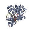
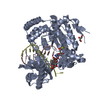
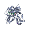
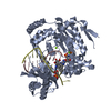
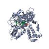
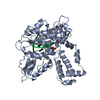
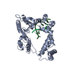
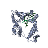
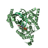
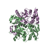
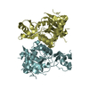
| Title | Crystal structure of D-alanine-D-alanine ligase from Acinetobacter baumannii |
|---|
 Components Components | D-alanine--D-alanine ligase |
|---|
 Keywords Keywords | LIGASE / D-alanine-D-alanine ligase / Acinetobacter baumannii / Apo structure / Drug target |
|---|
| Function / homology |  Function and homology information Function and homology information
D-alanine-D-alanine ligase / D-alanine-D-alanine ligase activity / peptidoglycan biosynthetic process / cell wall organization / regulation of cell shape / ATP binding / metal ion binding / cytosolSimilarity search - Function D-alanine--D-alanine ligase/VANA/B/C, conserved site / D-alanine--D-alanine ligase / D-alanine--D-alanine ligase, N-terminal domain / D-ala D-ala ligase N-terminus / D-alanine--D-alanine ligase signature 1. / D-alanine--D-alanine ligase signature 2. / D-alanine--D-alanine ligase, C-terminal / D-ala D-ala ligase C-terminus / Rossmann fold - #20 / ATP-grasp fold, A domain ...D-alanine--D-alanine ligase/VANA/B/C, conserved site / D-alanine--D-alanine ligase / D-alanine--D-alanine ligase, N-terminal domain / D-ala D-ala ligase N-terminus / D-alanine--D-alanine ligase signature 1. / D-alanine--D-alanine ligase signature 2. / D-alanine--D-alanine ligase, C-terminal / D-ala D-ala ligase C-terminus / Rossmann fold - #20 / ATP-grasp fold, A domain / ATP-grasp fold, B domain / Pre-ATP-grasp domain superfamily / ATP-grasp fold / ATP-grasp fold profile. / D-amino Acid Aminotransferase; Chain A, domain 1 / Dna Ligase; domain 1 / Rossmann fold / 2-Layer Sandwich / 3-Layer(aba) Sandwich / Alpha BetaSimilarity search - Domain/homology |
|---|
| Biological species |  Acinetobacter baumannii ACICU (bacteria) Acinetobacter baumannii ACICU (bacteria) |
|---|
| Method |  X-RAY DIFFRACTION / SYNCHROTRON / MOLECULAR REPLACEMENT / Resolution: 2.19 Å X-RAY DIFFRACTION / SYNCHROTRON / MOLECULAR REPLACEMENT / Resolution: 2.19 Å |
|---|
 Authors Authors | Huynh, K.H. / Hong, M.K. / Kang, L.W. |
|---|
 Citation Citation | Journal: J. Microbiol. / Year: 2015
Title: The crystal structure of the D-alanine-D-alanine ligase from Acinetobacter baumannii suggests a flexible conformational change in the central domain before nucleotide binding
Authors: Huynh, K.H. / Hong, M.K. / Lee, C. / Tran, H.T. / Lee, S.H. / Ahn, Y.J. / Cha, S.S. / Kang, L.W. |
|---|
| History | | Deposition | Aug 17, 2015 | Deposition site: RCSB / Processing site: PDBJ |
|---|
| Revision 1.0 | Aug 17, 2016 | Provider: repository / Type: Initial release |
|---|
| Revision 1.1 | Mar 20, 2024 | Group: Data collection / Database references / Derived calculations
Category: chem_comp_atom / chem_comp_bond ...chem_comp_atom / chem_comp_bond / citation / database_2 / pdbx_struct_oper_list
Item: _citation.country / _database_2.pdbx_DOI ..._citation.country / _database_2.pdbx_DOI / _database_2.pdbx_database_accession / _pdbx_struct_oper_list.symmetry_operation |
|---|
|
|---|
 Movie
Movie Controller

 Yorodumi
 Open data
 Basic information
 Structure visualization
 Downloads & links
 Other downloads
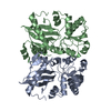

 PDBj
PDBj


 Assembly

 Mass: 18.015 Da / Num. of mol.: 199 / Source method: isolated from a natural source / Formula: H2O
 Sample preparation
 / Beamline: 5C (4A) / Wavelength: 0.9796 Å
 Processing