 Movie
Movie Controller

[English] 日本語
 Yorodumi
- PDB-1zqn: DNA POLYMERASE BETA (POL B) (E.C.2.7.7.7) COMPLEXED WITH SEVEN BA... -
+ Open data

- Basic information
| Entry | Database: PDB / ID: 1zqn | ||||||
|---|---|---|---|---|---|---|---|
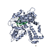
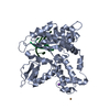
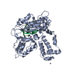
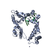
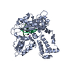
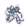
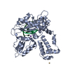
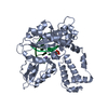
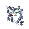
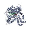
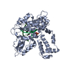
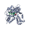
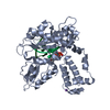
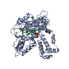
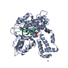
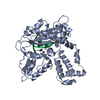
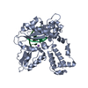
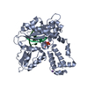
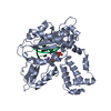
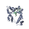
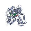
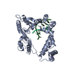
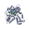
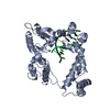
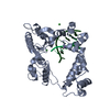
| Title | DNA POLYMERASE BETA (POL B) (E.C.2.7.7.7) COMPLEXED WITH SEVEN BASE PAIRS OF DNA; SOAKED IN THE PRESENCE OF BACL2 (15 MILLIMOLAR) AND NACL (15 MILLIMOLAR) | ||||||
 Components Components |
| ||||||
 Keywords Keywords | TRANSFERASE/DNA / DNA-DIRECTED DNA POLYMERASE / DNA REPLICATION / DNA REPAIR / NUCLEOTIDYLTRANSFERASE / COMPLEX (NUCLEOTIDYLTRANSFERASE-DNA / TRANSFERASE-DNA complex | ||||||
| Function / homology |  Function and homology information Function and homology informationResolution of AP sites via the single-nucleotide replacement pathway / immunoglobulin heavy chain V-D-J recombination / Resolution of AP sites via the multiple-nucleotide patch replacement pathway / Abasic sugar-phosphate removal via the single-nucleotide replacement pathway / APEX1-Independent Resolution of AP Sites via the Single Nucleotide Replacement Pathway / Lyases; Carbon-oxygen lyases; Other carbon-oxygen lyases / pyrimidine dimer repair / POLB-Dependent Long Patch Base Excision Repair / homeostasis of number of cells / PCNA-Dependent Long Patch Base Excision Repair ...Resolution of AP sites via the single-nucleotide replacement pathway / immunoglobulin heavy chain V-D-J recombination / Resolution of AP sites via the multiple-nucleotide patch replacement pathway / Abasic sugar-phosphate removal via the single-nucleotide replacement pathway / APEX1-Independent Resolution of AP Sites via the Single Nucleotide Replacement Pathway / Lyases; Carbon-oxygen lyases; Other carbon-oxygen lyases / pyrimidine dimer repair / POLB-Dependent Long Patch Base Excision Repair / homeostasis of number of cells / PCNA-Dependent Long Patch Base Excision Repair / 5'-deoxyribose-5-phosphate lyase activity / response to hyperoxia / lymph node development / salivary gland morphogenesis / somatic hypermutation of immunoglobulin genes / spleen development / base-excision repair, gap-filling / DNA-(apurinic or apyrimidinic site) endonuclease activity / class I DNA-(apurinic or apyrimidinic site) endonuclease activity / DNA-(apurinic or apyrimidinic site) lyase / response to gamma radiation / spindle microtubule / base-excision repair / DNA-templated DNA replication / double-strand break repair via nonhomologous end joining / intrinsic apoptotic signaling pathway in response to DNA damage / neuron apoptotic process / DNA-directed DNA polymerase / microtubule binding / in utero embryonic development / damaged DNA binding / microtubule / DNA-directed DNA polymerase activity / response to ethanol / lyase activity / Ub-specific processing proteases / inflammatory response / DNA repair / DNA damage response / enzyme binding / protein-containing complex / nucleoplasm / metal ion binding / nucleus / cytoplasm Similarity search - Function | ||||||
| Biological species |  Homo sapiens (human) Homo sapiens (human) | ||||||
| Method |  X-RAY DIFFRACTION / DIFFERENCE FOURIER / Resolution: 3 Å X-RAY DIFFRACTION / DIFFERENCE FOURIER / Resolution: 3 Å | ||||||
 Authors Authors | Pelletier, H. / Sawaya, M.R. | ||||||
 Citation Citation | Journal: Biochemistry / Year: 1996 Title: Characterization of the metal ion binding helix-hairpin-helix motifs in human DNA polymerase beta by X-ray structural analysis. Authors: Pelletier, H. / Sawaya, M.R. #1: Journal: To be PublishedTitle: Crystal Structures of Human DNA Polymerase Beta Complexed with Nicked and Gapped DNA Substrates Authors: Sawaya, M.R. / Rawson, T. / Wilson, S.H. / Kraut, J. / Pelletier, H. #2: Journal: To be PublishedTitle: The Role of Thumb Movement and Template Bending in Polymerase Fidelity Authors: Pelletier, H. #3: Journal: Biochemistry / Year: 1996Title: Crystal Structures of Human DNA Polymerase Beta Complexed with DNA; Implications for Catalytic Mechanism, Processivity, and Fidelity Authors: Pelletier, H. / Sawaya, M.R. / Wolfle, W. / Wilson, S.H. / Kraut, J. #4: Journal: Biochemistry / Year: 1996Title: A Structural Basis for Metal Ion Mutagenicity and Nucleotide Selectivity in Human DNA Polymerase Beta Authors: Pelletier, H. / Sawaya, M.R. / Wolfle, W. / Wilson, S.H. / Kraut, J. #6: Journal: Science / Year: 1994Title: Structures of Ternary Complexes of Rat DNA Polymerase Beta, a DNA Template- Primer, and ddCTP Authors: Pelletier, H. / Sawaya, M.R. / Kumar, A. / Wilson, S.H. / Kraut, J. #7: Journal: Science / Year: 1994Title: Crystal Structure of Rat DNA Polymerase Beta: Evidence for a Common Polymerase Mechanism Authors: Sawaya, M.R. / Pelletier, H. / Kumar, A. / Wilson, S.H. / Kraut, J. | ||||||
| History |
|
- Structure visualization
| Structure viewer | Molecule: MolmilJmol/JSmol |
|---|
- Downloads & links
-Download
| PDBx/mmCIF format | 1zqn.cif.gz | 90.8 KB | Display | PDBx/mmCIF format |
|---|---|---|---|---|
| PDB format | pdb1zqn.ent.gz | 70.2 KB | Display | PDB format |
| PDBx/mmJSON format | 1zqn.json.gz | Tree view | PDBx/mmJSON format | |
| Others |  Other downloads Other downloads |
-Validation report
| Arichive directory | https://data.pdbj.org/pub/pdb/validation_reports/zq/1zqnftp://data.pdbj.org/pub/pdb/validation_reports/zq/1zqn | HTTPS FTP |
|---|
-Related structure data
| Related structure data |  1nomC  1zqaC 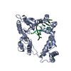 1zqbC 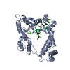 1zqcC 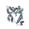 1zqdC  1zqeC 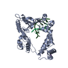 1zqfC  1zqgSC  1zqhC  1zqiC 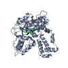 1zqjC  1zqkC  1zqlC  1zqmC  1zqoC  1zqpC  1zqqC  1zqrC  1zqsC  1zquC  1zqvC  1zqwC  1zqxC  1zqyC  1zqzC 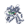 8icaC  8icbC 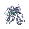 8iccC 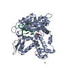 8iceC  8icfC  8icgC  8ichC  8iciC  8iczC  9icaC  9icbC  9iccC  9iceC  9icgC  9ichC  9iciC 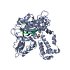 9icjC  9iclC C: citing same article ( S: Starting model for refinement |
|---|---|
| Similar structure data |


-Links
 PDBj
PDBj


- Assembly
| Deposited unit | 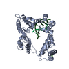
| ||||||||
|---|---|---|---|---|---|---|---|---|---|
| 1 |
| ||||||||
| Unit cell |
|
-Components
| #1: DNA chain | Mass: 2434.643 Da / Num. of mol.: 1 / Source method: obtained synthetically | ||||
|---|---|---|---|---|---|
| #2: DNA chain | Mass: 2112.422 Da / Num. of mol.: 1 / Source method: obtained synthetically | ||||
| #3: Protein | Mass: 38241.672 Da / Num. of mol.: 1 Source method: isolated from a genetically manipulated source Source: (gene. exp.) Homo sapiens (human) / Production host:  | ||||
| #4: Chemical |   Mass: 137.327 Da / Num. of mol.: 2 / Source method: obtained synthetically / Formula: Ba Mass: 137.327 Da / Num. of mol.: 2 / Source method: obtained synthetically / Formula: Ba#5: Water | ChemComp-HOH / |  Mass: 18.015 Da / Num. of mol.: 144 / Source method: isolated from a natural source / Formula: H2O Mass: 18.015 Da / Num. of mol.: 144 / Source method: isolated from a natural source / Formula: H2OCompound details | A POSSIBLE PHYSIOLOGICAL SUBSTRATE FOR POL BETA IS A DNA GAP, WHERE THE LENGTH OF THE GAP CAN RANGE ...A POSSIBLE PHYSIOLOGI | |
-Experimental details
-Experiment
| Experiment | Method: X-RAY DIFFRACTION / Number of used crystals: 1 |
|---|
- Sample preparation
| Crystal | Density Matthews: 3 Å3/Da / Density % sol: 58.4 % |
|---|---|
| Crystal grow | pH: 6.5 Details: THIS ENTRY DESCRIBES THE STRUCTURE THAT RESULTED WHEN A COCRYSTAL OF HUMAN DNA POLYMERASE BETA COMPLEXED WITH 7 BASE PAIRS OF DNA (SEE ENTRY 9ICJ AND REFERENCE 1) HAD BEEN SOAKED IN THE ...Details: THIS ENTRY DESCRIBES THE STRUCTURE THAT RESULTED WHEN A COCRYSTAL OF HUMAN DNA POLYMERASE BETA COMPLEXED WITH 7 BASE PAIRS OF DNA (SEE ENTRY 9ICJ AND REFERENCE 1) HAD BEEN SOAKED IN THE FOLLOWING SOLUTION FOR 3 DAYS: PEG 3350, 16% IMIDAZOLE, 100 MILLIMOLAR, PH 6.5 BACL2, 15 MILLIMOLAR NACL, 15 MILLIMOLAR SEE REFERENCE 3 FOR DETAILS CONCERNING EXPERIMENTAL PROCEDURES, RESULTS, AND DISCUSSION FOR THIS STRUCTURE. |
-Data collection
| Diffraction | Mean temperature: 298 K |
|---|---|
| Diffraction source | Source: ROTATING ANODE / Type: RIGAKU RU200 |
| Detector | Type: SDMS / Detector: AREA DETECTOR / Date: Mar 25, 1996 |
| Radiation | Monochromator: GRAPHITE / Monochromatic (M) / Laue (L): M / Scattering type: x-ray |
| Radiation wavelength | Relative weight: 1 |
| Reflection | Resolution: 3→20 Å / Num. obs: 9206 / % possible obs: 89 % / Observed criterion σ(I): 0 / Redundancy: 1.9 % / Rsym value: 0.076 |
| Reflection shell | Resolution: 3→3.1 Å / Redundancy: 1.4 % / Rsym value: 0.157 / % possible all: 88 |
- Processing
| Software |
| ||||||||||||||||||||||||||||||||||||||||||||||||||
|---|---|---|---|---|---|---|---|---|---|---|---|---|---|---|---|---|---|---|---|---|---|---|---|---|---|---|---|---|---|---|---|---|---|---|---|---|---|---|---|---|---|---|---|---|---|---|---|---|---|---|---|
| Refinement | Method to determine structure: DIFFERENCE FOURIER Starting model: 1ZQG Resolution: 3→20 Å / σ(F): 0 / Stereochemistry target values: STANDARD TNT /
| ||||||||||||||||||||||||||||||||||||||||||||||||||
| Solvent computation | Solvent model: MOEWS / Bsol: 202.2 Å2 / ksol: 0.8 e/Å3 | ||||||||||||||||||||||||||||||||||||||||||||||||||
| Refinement step | Cycle: LAST / Resolution: 3→20 Å
| ||||||||||||||||||||||||||||||||||||||||||||||||||
| Refine LS restraints |
|
