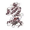
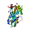
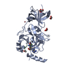
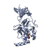
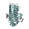
| Deposited unit | A: Protein kinase
B: Protein kinase
C: Protein kinase
D: Protein kinase
E: Protein kinase
F: Protein kinase
hetero molecules
| Theoretical mass | Number of molelcules |
|---|
| Total (without water) | 202,313 | 15 |
|---|
| Polymers | 198,915 | 6 |
|---|
| Non-polymers | 3,398 | 9 |
|---|
| Water | 0 | 0 |
|---|
|
|---|
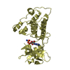
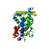
| 1 | A: Protein kinase
hetero molecules
| Theoretical mass | Number of molelcules |
|---|
| Total (without water) | 33,779 | 3 |
|---|
| Polymers | 33,153 | 1 |
|---|
| Non-polymers | 626 | 2 |
|---|
| Water | 0 | |
|---|
| Type | Name | Symmetry operation | Number |
|---|
| identity operation | 1_555 | x,y,z | 1 |
|
|---|
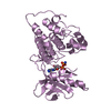
| 2 | B: Protein kinase
hetero molecules
| Theoretical mass | Number of molelcules |
|---|
| Total (without water) | 33,779 | 3 |
|---|
| Polymers | 33,153 | 1 |
|---|
| Non-polymers | 626 | 2 |
|---|
| Water | 0 | |
|---|
| Type | Name | Symmetry operation | Number |
|---|
| identity operation | 1_555 | x,y,z | 1 |
|
|---|
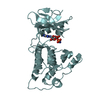
| 3 | C: Protein kinase
hetero molecules
| Theoretical mass | Number of molelcules |
|---|
| Total (without water) | 33,779 | 3 |
|---|
| Polymers | 33,153 | 1 |
|---|
| Non-polymers | 626 | 2 |
|---|
| Water | 0 | |
|---|
| Type | Name | Symmetry operation | Number |
|---|
| identity operation | 1_555 | x,y,z | 1 |
|
|---|
| 4 | D: Protein kinase
hetero molecules
| Theoretical mass | Number of molelcules |
|---|
| Total (without water) | 33,659 | 2 |
|---|
| Polymers | 33,153 | 1 |
|---|
| Non-polymers | 506 | 1 |
|---|
| Water | 0 | |
|---|
| Type | Name | Symmetry operation | Number |
|---|
| identity operation | 1_555 | x,y,z | 1 |
|
|---|
| 5 | E: Protein kinase
hetero molecules
| Theoretical mass | Number of molelcules |
|---|
| Total (without water) | 33,659 | 2 |
|---|
| Polymers | 33,153 | 1 |
|---|
| Non-polymers | 506 | 1 |
|---|
| Water | 0 | |
|---|
| Type | Name | Symmetry operation | Number |
|---|
| identity operation | 1_555 | x,y,z | 1 |
|
|---|
| 6 | F: Protein kinase
hetero molecules
| Theoretical mass | Number of molelcules |
|---|
| Total (without water) | 33,659 | 2 |
|---|
| Polymers | 33,153 | 1 |
|---|
| Non-polymers | 506 | 1 |
|---|
| Water | 0 | |
|---|
| Type | Name | Symmetry operation | Number |
|---|
| identity operation | 1_555 | x,y,z | 1 |
|
|---|
| Unit cell | | Length a, b, c (Å) | 221.510, 127.550, 70.280 |
|---|
| Angle α, β, γ (deg.) | 90.00, 89.96, 90.00 |
|---|
| Int Tables number | 5 |
|---|
| Space group name H-M | C121 |
|---|
|
|---|
| Noncrystallographic symmetry (NCS) | NCS domain: | ID | Ens-ID |
|---|
| 1 | 1 | | 2 | 1 | | 3 | 1 | | 1 | 2 | | 2 | 2 | | 3 | 2 | | 1 | 3 | | 2 | 3 | | 3 | 3 | | 1 | 4 | | 2 | 4 | | 3 | 4 | | 1 | 5 | | 2 | 5 | | 3 | 5 | | 1 | 6 | | 2 | 6 | | 3 | 6 | | 1 | 7 | | 2 | 7 | | 3 | 7 | | 1 | 8 | | 2 | 8 | | 3 | 8 | | 1 | 9 | | 2 | 9 | | 3 | 9 | | 1 | 10 | | 2 | 10 | | 3 | 10 | | 1 | 11 | | 2 | 11 | | 3 | 11 | | 1 | 12 | | 2 | 12 | | 3 | 12 | | 1 | 13 | | 2 | 13 | | 3 | 13 | | 1 | 14 | | 2 | 14 | | 3 | 14 | | 1 | 15 | | 2 | 15 | | 3 | 15 | | 1 | 16 | | 2 | 16 | | 3 | 16 | | 1 | 17 | | 2 | 17 | | 3 | 17 | | 1 | 18 | | 2 | 18 | | 3 | 18 |
NCS domain segments: | Dom-ID | Component-ID | Ens-ID | Selection details |
|---|
| 1 | 1 | 1 | chain A and (resseq 7:16 )| 2 | 1 | 1 | chain B and (resseq 7:16 )| 3 | 1 | 1 | chain C and (resseq 7:16 )| 1 | 1 | 2 | chain D and (resseq 7:16 )| 2 | 1 | 2 | chain E and (resseq 7:16 )| 3 | 1 | 2 | chain F and (resseq 7:16 )| 1 | 1 | 3 | chain A and (resseq 22:29 )| 2 | 1 | 3 | chain B and (resseq 22:29 )| 3 | 1 | 3 | chain C and (resseq 22:29 )| 1 | 1 | 4 | chain A and (resseq 35:41 )| 2 | 1 | 4 | chain B and (resseq 35:41 )| 3 | 1 | 4 | chain C and (resseq 35:41 )| 1 | 1 | 5 | chain D and (resseq 22:29 )| 2 | 1 | 5 | chain E and (resseq 22:29 )| 3 | 1 | 5 | chain F and (resseq 22:29 )| 1 | 1 | 6 | chain D and (resseq 35:41 )| 2 | 1 | 6 | chain E and (resseq 35:41 )| 3 | 1 | 6 | chain F and ( | | | | | | | | | | | | | | | | | |
|
|---|
 Movie
Movie Controller

 Yorodumi
 Open data
 Basic information
 Components
 Keywords
 Function and homology information
 Staphylococcus aureus subsp. aureus (bacteria)
 X-RAY DIFFRACTION /
  Authors
 Citation
 Structure visualization
 Downloads & links
 Other downloads
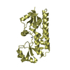


 PDBj
PDBj


 Assembly