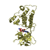
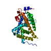
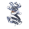
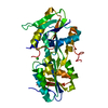
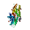
- PDB-5gt4: Crystal structure of the human vitamin D receptor ligand binding ... -
+
Open data
ID or keywords:
Loading...
-
Basic information
Entry
Database: PDB / ID: 5gt4
Title
Crystal structure of the human vitamin D receptor ligand binding domain complexed with (1R,2S,3R,5Z,7E,14beta,17alpha)-2-cyanopropoxy-9,10-secocholesta-5,7,10-triene-1,3,25-triol
Components
Vitamin D3 receptor
Keywords
TRANSCRIPTION / nuclear hormone receptor full agonist ligand complex
Function / homology
Function and homology information
apoptotic process involved in mammary gland involution / response to bile acid / Vitamin D (calciferol) metabolism / positive regulation of apoptotic process involved in mammary gland involution / calcitriol binding / lithocholic acid binding / nuclear receptor-mediated bile acid signaling pathway / bile acid nuclear receptor activity / positive regulation of keratinocyte differentiation / vitamin D binding ...apoptotic process involved in mammary gland involution / response to bile acid / Vitamin D (calciferol) metabolism / positive regulation of apoptotic process involved in mammary gland involution / calcitriol binding / lithocholic acid binding / nuclear receptor-mediated bile acid signaling pathway / bile acid nuclear receptor activity / positive regulation of keratinocyte differentiation / vitamin D binding / vitamin D receptor signaling pathway / positive regulation of vitamin D receptor signaling pathway / phosphate ion transmembrane transport / intestinal absorption / mammary gland branching involved in pregnancy / decidualization / negative regulation of keratinocyte proliferation / positive regulation of bone mineralization / nuclear retinoid X receptor binding / retinoic acid receptor signaling pathway / intracellular receptor signaling pathway / lactation / skeletal system development / SUMOylation of intracellular receptors / Nuclear Receptor transcription pathway / mRNA transcription by RNA polymerase II / cell morphogenesis / nuclear receptor activity / RNA polymerase II transcription regulator complex / intracellular calcium ion homeostasis / calcium ion transport / DNA-binding transcription factor activity, RNA polymerase II-specific / cell differentiation / signaling receptor complex / RNA polymerase II cis-regulatory region sequence-specific DNA binding / negative regulation of cell population proliferation / negative regulation of DNA-templated transcription / positive regulation of gene expression / chromatin / negative regulation of transcription by RNA polymerase II / positive regulation of transcription by RNA polymerase II / DNA binding / zinc ion binding / nucleoplasm / nucleus / cytosol Similarity search - Function
Journal: To Be Published Title: Crystal structure of the human vitamin D receptor ligand binding domain complexed with (1R,2S,3R,5Z,7E,14beta,17alpha)-2-cyanopropoxy-9,10-secocholesta-5,7,10-triene-1,3,25-triol Authors: Takimoto-Kamimura, M.
Resolution: 1.83→48.27 Å / Cor.coef. Fo:Fc: 0.945 / Cor.coef. Fo:Fc free: 0.925 / SU B: 2.414 / SU ML: 0.076 / SU R Cruickshank DPI: 0.1239 / Cross valid method: THROUGHOUT / σ(F): 0 / ESU R: 0.124 / ESU R Free: 0.116 Details: The authors state they removed the long loop 165-215 for the crystallization with direct lincage with Gly 164 and Ser 216, so this region recieved quite large steric hindrance and resuled ...Details: The authors state they removed the long loop 165-215 for the crystallization with direct lincage with Gly 164 and Ser 216, so this region recieved quite large steric hindrance and resuled abnormal bond length as usual, and that the bond is existing in real but the bond distance is abnormal by the disorder by the steric hindrances.
Rfactor
Num. reflection
% reflection
Selection details
Rfree
0.2171
1394
5 %
RANDOM
Rwork
0.1904
-
-
-
obs
0.1918
26427
99.76 %
-
Solvent computation
Ion probe radii: 0.8 Å / Shrinkage radii: 0.8 Å / VDW probe radii: 1.2 Å
In the structure databanks used in Yorodumi, some data are registered as the other names, "COVID-19 virus" and "2019-nCoV". Here are the details of the virus and the list of structure data.
Jan 31, 2019. EMDB accession codes are about to change! (news from PDBe EMDB page)
EMDB accession codes are about to change! (news from PDBe EMDB page)
The allocation of 4 digits for EMDB accession codes will soon come to an end. Whilst these codes will remain in use, new EMDB accession codes will include an additional digit and will expand incrementally as the available range of codes is exhausted. The current 4-digit format prefixed with “EMD-” (i.e. EMD-XXXX) will advance to a 5-digit format (i.e. EMD-XXXXX), and so on. It is currently estimated that the 4-digit codes will be depleted around Spring 2019, at which point the 5-digit format will come into force.
The EM Navigator/Yorodumi systems omit the EMD- prefix.
Related info.:Q: What is EMD? / ID/Accession-code notation in Yorodumi/EM Navigator
Yorodumi is a browser for structure data from EMDB, PDB, SASBDB, etc.
This page is also the successor to EM Navigator detail page, and also detail information page/front-end page for Omokage search.
The word "yorodu" (or yorozu) is an old Japanese word meaning "ten thousand". "mi" (miru) is to see.
Related info.:EMDB / PDB / SASBDB / Comparison of 3 databanks / Yorodumi Search / Aug 31, 2016. New EM Navigator & Yorodumi / Yorodumi Papers / Jmol/JSmol / Function and homology information / Changes in new EM Navigator and Yorodumi
 Movie
Movie Controller

 Yorodumi
 Open data
 Basic information
 Components
 Keywords
 Function and homology information
 Homo sapiens (human)
 X-RAY DIFFRACTION /
  Authors
 Citation
 Structure visualization
 Downloads & links
 Other downloads
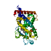


 PDBj
PDBj

 Assembly
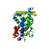

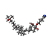
 Mass: 499.725 Da / Num. of mol.: 1 / Source method: obtained synthetically / Formula: C31H49NO4
 Mass: 18.015 Da / Num. of mol.: 71 / Source method: isolated from a natural source / Formula: H2O
 Sample preparation
 / Beamline: MX2 / Wavelength: 0.9537 Å
 Processing