 Movie
Movie Controller


+ Open data

- Basic information
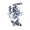
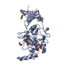
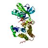
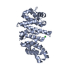
| Entry | Database: PDB / ID: 3m59 | ||||||
|---|---|---|---|---|---|---|---|
| Title | SET7/9 Y245A in complex with TAF10-K189me2 peptide and AdoHcy | ||||||
 Components Components |
| ||||||
 Keywords Keywords | TRANSFERASE / TERNARY COMPLEX / SET DOMAIN / METHYLTRANSFERASE / S-ADENOSYL-L-HOMOCYSTEINE / TAF10 PEPTIDE / N-DIMETHYLLYSINE / Chromatin regulator / Chromosomal protein / Nucleus / S-adenosyl-L-methionine / Transcription / Transcription regulation | ||||||
| Function / homology |  Function and homology information Function and homology informationSAGA complex assembly / lateral mesodermal cell differentiation / allantois development / peptidyl-lysine monomethylation / heterochromatin organization / peptidyl-lysine dimethylation / transcription factor TFTC complex / [histone H3]-lysine4 N-methyltransferase / histone H3K4 monomethyltransferase activity / SAGA complex ...SAGA complex assembly / lateral mesodermal cell differentiation / allantois development / peptidyl-lysine monomethylation / heterochromatin organization / peptidyl-lysine dimethylation / transcription factor TFTC complex / [histone H3]-lysine4 N-methyltransferase / histone H3K4 monomethyltransferase activity / SAGA complex / protein-lysine N-methyltransferase activity / RNA polymerase binding / transcription preinitiation complex / histone H3 methyltransferase activity / histone methyltransferase activity / transcription factor TFIID complex / RNA polymerase II general transcription initiation factor activity / limb development / HIV Transcription Initiation / RNA Polymerase II HIV Promoter Escape / Transcription of the HIV genome / RNA Polymerase II Promoter Escape / RNA Polymerase II Transcription Pre-Initiation And Promoter Opening / RNA Polymerase II Transcription Initiation / RNA Polymerase II Transcription Initiation And Promoter Clearance / regulation of RNA splicing / somitogenesis / embryonic placenta development / positive regulation of transcription initiation by RNA polymerase II / RNA polymerase II preinitiation complex assembly / regulation of DNA repair / RNA Polymerase II Pre-transcription Events / nuclear estrogen receptor binding / promoter-specific chromatin binding / DNA-templated transcription initiation / transcription initiation at RNA polymerase II promoter / PKMTs methylate histone lysines / mRNA transcription by RNA polymerase II / p53 binding / transcription by RNA polymerase II / chromosome / HATs acetylate histones / chromatin organization / Regulation of TP53 Activity through Phosphorylation / Ub-specific processing proteases / chromatin remodeling / regulation of transcription by RNA polymerase II / DNA damage response / regulation of DNA-templated transcription / nucleolus / positive regulation of DNA-templated transcription / perinuclear region of cytoplasm / enzyme binding / DNA-templated transcription / DNA binding / nucleoplasm / identical protein binding / nucleus / cytoplasm Similarity search - Function | ||||||
| Biological species |  Homo sapiens (human) Homo sapiens (human) | ||||||
| Method |  X-RAY DIFFRACTION / SYNCHROTRON / MOLECULAR REPLACEMENT / molecular replacement / Resolution: 1.7 Å X-RAY DIFFRACTION / SYNCHROTRON / MOLECULAR REPLACEMENT / molecular replacement / Resolution: 1.7 Å | ||||||
 Authors Authors | Del Rizzo, P.A. / Couture, J.-F. / Roiko, M.S. / Strunk, B.S. / Brunzelle, J.S. / Dirk, L.M. / Houtz, R.L. / Trievel, R.C. | ||||||
 Citation Citation | Journal: J.Biol.Chem. / Year: 2010 Title: SET7/9 catalytic mutants reveal the role of active site water molecules in lysine multiple methylation. Authors: Del Rizzo, P.A. / Couture, J.F. / Dirk, L.M. / Strunk, B.S. / Roiko, M.S. / Brunzelle, J.S. / Houtz, R.L. / Trievel, R.C. | ||||||
| History |
|
- Structure visualization
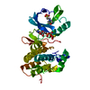
| Structure viewer | Molecule: MolmilJmol/JSmol |
|---|
- Downloads & links
-Download
| PDBx/mmCIF format | 3m59.cif.gz | 75.3 KB | Display | PDBx/mmCIF format |
|---|---|---|---|---|
| PDB format | pdb3m59.ent.gz | 53.1 KB | Display | PDB format |
| PDBx/mmJSON format | 3m59.json.gz | Tree view | PDBx/mmJSON format | |
| Others |  Other downloads Other downloads |
-Validation report
| Arichive directory | https://data.pdbj.org/pub/pdb/validation_reports/m5/3m59ftp://data.pdbj.org/pub/pdb/validation_reports/m5/3m59 | HTTPS FTP |
|---|
-Related structure data
| Related structure data | 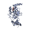 3m53C 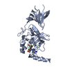 3m54C 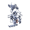 3m55C 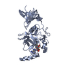 3m56C 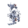 3m57C 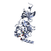 3m58C 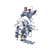 3m5aC 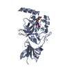 2f69S S: Starting model for refinement C: citing same article ( |
|---|---|
| Similar structure data |


-Links
 PDBj
PDBj


- Assembly
| Deposited unit | 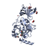
| |||||||||
|---|---|---|---|---|---|---|---|---|---|---|
| 1 |
| |||||||||
| Unit cell |
| |||||||||
| Components on special symmetry positions |
|
-Components
-Protein / Protein/peptide , 2 types, 2 molecules AB
| #1: Protein | Mass: 28951.236 Da / Num. of mol.: 1 / Fragment: UNP residues 110-366 / Mutation: Y245A Source method: isolated from a genetically manipulated source Source: (gene. exp.) Homo sapiens (human) / Strain: 9606 / Gene: KIAA1717, KMT7, SET7, SET9, SETD7 / Plasmid: pHIS2 / Production host:  References: UniProt: Q8WTS6, histone-lysine N-methyltransferase |
|---|---|
| #2: Protein/peptide | Mass: 1282.511 Da / Num. of mol.: 1 / Source method: obtained synthetically / Source: (synth.) Homo sapiens (human) / References: UniProt: Q12962*PLUS |
-Non-polymers , 4 types, 245 molecules 


| #3: Chemical | ChemComp-SAH /  Type: L-peptide linking / Mass: 384.411 Da / Num. of mol.: 1 / Source method: obtained synthetically / Formula: C14H20N6O5S Type: L-peptide linking / Mass: 384.411 Da / Num. of mol.: 1 / Source method: obtained synthetically / Formula: C14H20N6O5S | ||||
|---|---|---|---|---|---|
| #4: Chemical |  Mass: 58.933 Da / Num. of mol.: 3 / Source method: obtained synthetically / Formula: Co Mass: 58.933 Da / Num. of mol.: 3 / Source method: obtained synthetically / Formula: Co#5: Chemical | ChemComp-GOL /  Mass: 92.094 Da / Num. of mol.: 4 / Source method: obtained synthetically / Formula: C3H8O3 Mass: 92.094 Da / Num. of mol.: 4 / Source method: obtained synthetically / Formula: C3H8O3#6: Water | ChemComp-HOH / | Mass: 18.015 Da / Num. of mol.: 237 / Source method: isolated from a natural source / Formula: H2O |
-Experimental details
-Experiment
| Experiment | Method: X-RAY DIFFRACTION / Number of used crystals: 1 |
|---|
- Sample preparation
| Crystal | Density Matthews: 3.19 Å3/Da / Density % sol: 61.39 % |
|---|---|
| Crystal grow | Temperature: 293 K / Method: vapor diffusion, hanging drop / pH: 6.4 Details: 1.9 M Ammonium Sulfate, 0.1 M Bis-Tris pH 6.4, 0.005 M CoCl2, VAPOR DIFFUSION, HANGING DROP, temperature 293K |
-Data collection
| Diffraction | Mean temperature: 100 K | |||||||||||||||||||||||||||||||||||||||||||||||||||||||||||||||||||||||||||||
|---|---|---|---|---|---|---|---|---|---|---|---|---|---|---|---|---|---|---|---|---|---|---|---|---|---|---|---|---|---|---|---|---|---|---|---|---|---|---|---|---|---|---|---|---|---|---|---|---|---|---|---|---|---|---|---|---|---|---|---|---|---|---|---|---|---|---|---|---|---|---|---|---|---|---|---|---|---|---|
| Diffraction source | Source: SYNCHROTRON / Site: APS  / Beamline: 21-ID-G / Wavelength: 0.9786 Å / Beamline: 21-ID-G / Wavelength: 0.9786 Å | |||||||||||||||||||||||||||||||||||||||||||||||||||||||||||||||||||||||||||||
| Detector | Type: MARMOSAIC 300 mm CCD / Detector: CCD / Date: Aug 2, 2008 | |||||||||||||||||||||||||||||||||||||||||||||||||||||||||||||||||||||||||||||
| Radiation | Monochromator: C(111) / Protocol: SINGLE WAVELENGTH / Monochromatic (M) / Laue (L): M / Scattering type: x-ray | |||||||||||||||||||||||||||||||||||||||||||||||||||||||||||||||||||||||||||||
| Radiation wavelength | Wavelength: 0.9786 Å / Relative weight: 1 | |||||||||||||||||||||||||||||||||||||||||||||||||||||||||||||||||||||||||||||
| Reflection | Resolution: 1.7→40 Å / Num. obs: 40597 / % possible obs: 95 % / Redundancy: 6 % / Rmerge(I) obs: 0.055 / Χ2: 1.394 / Net I/σ(I): 15.1 | |||||||||||||||||||||||||||||||||||||||||||||||||||||||||||||||||||||||||||||
| Reflection shell |
|
-Phasing
| Phasing | Method: molecular replacement | |||||||||
|---|---|---|---|---|---|---|---|---|---|---|
| Phasing MR |
|
- Processing
| Software |
| |||||||||||||||||||||||||||||||||||||||||||||||||||||||||||||||||
|---|---|---|---|---|---|---|---|---|---|---|---|---|---|---|---|---|---|---|---|---|---|---|---|---|---|---|---|---|---|---|---|---|---|---|---|---|---|---|---|---|---|---|---|---|---|---|---|---|---|---|---|---|---|---|---|---|---|---|---|---|---|---|---|---|---|---|
| Refinement | Method to determine structure: MOLECULAR REPLACEMENT Starting model: PDB entry 2F69 Resolution: 1.7→36.16 Å / Cor.coef. Fo:Fc: 0.963 / Cor.coef. Fo:Fc free: 0.958 / WRfactor Rfree: 0.211 / WRfactor Rwork: 0.188 / Occupancy max: 1 / Occupancy min: 0.3 / FOM work R set: 0.872 / SU B: 1.733 / SU ML: 0.057 / SU R Cruickshank DPI: 0.092 / SU Rfree: 0.089 / Cross valid method: THROUGHOUT / σ(F): 0 / ESU R: 0.092 / ESU R Free: 0.089 / Stereochemistry target values: MAXIMUM LIKELIHOOD Details: HYDROGENS HAVE BEEN ADDED IN THE RIDING POSITIONS U VALUES: REFINED INDIVIDUALLY
| |||||||||||||||||||||||||||||||||||||||||||||||||||||||||||||||||
| Solvent computation | Ion probe radii: 0.8 Å / Shrinkage radii: 0.8 Å / VDW probe radii: 1.2 Å / Solvent model: MASK | |||||||||||||||||||||||||||||||||||||||||||||||||||||||||||||||||
| Displacement parameters | Biso max: 102.66 Å2 / Biso mean: 27.057 Å2 / Biso min: 11.76 Å2
| |||||||||||||||||||||||||||||||||||||||||||||||||||||||||||||||||
| Refinement step | Cycle: LAST / Resolution: 1.7→36.16 Å
| |||||||||||||||||||||||||||||||||||||||||||||||||||||||||||||||||
| Refine LS restraints |
| |||||||||||||||||||||||||||||||||||||||||||||||||||||||||||||||||
| LS refinement shell | Resolution: 1.7→1.747 Å / Total num. of bins used: 20
|
