 Movie
Movie Controller

[English] 日本語
 Yorodumi
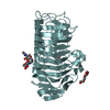
- PDB-6kvh: The mutant crystal structure of endo-polygalacturonase (T284A) fr... -
+ Open data

- Basic information
| Entry | Database: PDB / ID: 6kvh | ||||||
|---|---|---|---|---|---|---|---|
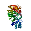
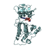
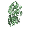
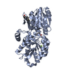
| Title | The mutant crystal structure of endo-polygalacturonase (T284A) from Talaromyces leycettanus JCM 12802 | ||||||
 Components Components | endo-polygalacturonase | ||||||
 Keywords Keywords | HYDROLASE / endo-polygalacturonase | ||||||
| Function / homology | alpha-D-mannopyranose Function and homology information Function and homology information | ||||||
| Biological species |  Talaromyces leycettanus (fungus) Talaromyces leycettanus (fungus) | ||||||
| Method |  X-RAY DIFFRACTION / SYNCHROTRON / MOLECULAR REPLACEMENT / Resolution: 1.2 Å X-RAY DIFFRACTION / SYNCHROTRON / MOLECULAR REPLACEMENT / Resolution: 1.2 Å | ||||||
 Authors Authors | Tu, T. / Wang, Z. / Luo, H. / Yao, B. | ||||||
| Funding support |  China, 1items China, 1items
| ||||||
 Citation Citation | Journal: J.Agric.Food Chem. / Year: 2021 Title: Structural Insights into the Mechanisms Underlying the Kinetic Stability of GH28 Endo-Polygalacturonase. Authors: Tu, T. / Wang, Z. / Luo, Y. / Li, Y. / Su, X. / Wang, Y. / Zhang, J. / Rouvinen, J. / Yao, B. / Hakulinen, N. / Luo, H. | ||||||
| History |
|
- Structure visualization
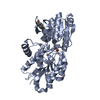
| Structure viewer | Molecule: MolmilJmol/JSmol |
|---|
- Downloads & links
-Download
| PDBx/mmCIF format | 6kvh.cif.gz | 187.9 KB | Display | PDBx/mmCIF format |
|---|---|---|---|---|
| PDB format | pdb6kvh.ent.gz | 122.8 KB | Display | PDB format |
| PDBx/mmJSON format | 6kvh.json.gz | Tree view | PDBx/mmJSON format | |
| Others |  Other downloads Other downloads |
-Validation report
| Arichive directory | https://data.pdbj.org/pub/pdb/validation_reports/kv/6kvhftp://data.pdbj.org/pub/pdb/validation_reports/kv/6kvh | HTTPS FTP |
|---|
-Related structure data
| Related structure data |  6kveSC S: Starting model for refinement C: citing same article ( |
|---|---|
| Similar structure data |


-Links
 PDBj
PDBj


- Assembly
| Deposited unit | 
| ||||||||||||
|---|---|---|---|---|---|---|---|---|---|---|---|---|---|
| 1 |
| ||||||||||||
| Unit cell |
|
-Components
| #1: Protein | Mass: 35208.250 Da / Num. of mol.: 1 Source method: isolated from a genetically manipulated source Source: (gene. exp.) Talaromyces leycettanus (fungus) / Strain: JCM 12802 / Production host: Komagataella phaffii GS115 (fungus) |
|---|---|
| #2: Sugar | ChemComp-NAG /   Type: D-saccharide, beta linking / Mass: 221.208 Da / Num. of mol.: 1 Type: D-saccharide, beta linking / Mass: 221.208 Da / Num. of mol.: 1Source method: isolated from a genetically manipulated source Formula: C8H15NO6 / Feature type: SUBJECT OF INVESTIGATION |
| #3: Sugar | ChemComp-MAN /   Type: D-saccharide, alpha linking / Mass: 180.156 Da / Num. of mol.: 1 Type: D-saccharide, alpha linking / Mass: 180.156 Da / Num. of mol.: 1Source method: isolated from a genetically manipulated source Formula: C6H12O6 / Feature type: SUBJECT OF INVESTIGATION |
| #4: Water | ChemComp-HOH /  Mass: 18.015 Da / Num. of mol.: 392 / Source method: isolated from a natural source / Formula: H2O Mass: 18.015 Da / Num. of mol.: 392 / Source method: isolated from a natural source / Formula: H2O |
| Has ligand of interest | Y |
| Has protein modification | Y |
-Experimental details
-Experiment
| Experiment | Method: X-RAY DIFFRACTION / Number of used crystals: 1 |
|---|
- Sample preparation
| Crystal | Density Matthews: 1.95 Å3/Da / Density % sol: 37.05 % |
|---|---|
| Crystal grow | Temperature: 293 K / Method: vapor diffusion, hanging drop / pH: 3.5 Details: 0.1 M citric acid (pH 3.5), 25% (w/v) polyethylene glycol 3350 |
-Data collection
| Diffraction | Mean temperature: 100 K / Serial crystal experiment: N |
|---|---|
| Diffraction source | Source: SYNCHROTRON / Site: SSRF / Beamline: BL17U / Wavelength: 1 Å |
| Detector | Type: DECTRIS PILATUS3 6M / Detector: PIXEL / Date: May 20, 2019 |
| Radiation | Protocol: SINGLE WAVELENGTH / Monochromatic (M) / Laue (L): M / Scattering type: x-ray |
| Radiation wavelength | Wavelength: 1 Å / Relative weight: 1 |
| Reflection | Resolution: 1.198→25.93 Å / Num. obs: 80946 / % possible obs: 94.95 % / Redundancy: 1.3 % / Biso Wilson estimate: 15.24 Å2 / CC1/2: 1 / Rmerge(I) obs: 0.01 / Rpim(I) all: 0.01 / Rrim(I) all: 0.01146 / Net I/σ(I): 15.72 |
| Reflection shell | Resolution: 1.198→1.241 Å / Redundancy: 1 % / Rmerge(I) obs: 0.01 / Mean I/σ(I) obs: 3.94 / Num. unique obs: 6194 / CC1/2: 1 / Rpim(I) all: 0.01 / Rrim(I) all: 0.01 / % possible all: 73.61 |
- Processing
| Software |
| ||||||||||||||||||||||||||||||||||||||||||||||||||||||||||||||||||||||||||||||||||||||||||||||||||||||||||||||||||||||||||||||||||||||||||||||||||||||||||||||||||||||||||||||||||||||||||||||||||||||||||||||||||
|---|---|---|---|---|---|---|---|---|---|---|---|---|---|---|---|---|---|---|---|---|---|---|---|---|---|---|---|---|---|---|---|---|---|---|---|---|---|---|---|---|---|---|---|---|---|---|---|---|---|---|---|---|---|---|---|---|---|---|---|---|---|---|---|---|---|---|---|---|---|---|---|---|---|---|---|---|---|---|---|---|---|---|---|---|---|---|---|---|---|---|---|---|---|---|---|---|---|---|---|---|---|---|---|---|---|---|---|---|---|---|---|---|---|---|---|---|---|---|---|---|---|---|---|---|---|---|---|---|---|---|---|---|---|---|---|---|---|---|---|---|---|---|---|---|---|---|---|---|---|---|---|---|---|---|---|---|---|---|---|---|---|---|---|---|---|---|---|---|---|---|---|---|---|---|---|---|---|---|---|---|---|---|---|---|---|---|---|---|---|---|---|---|---|---|---|---|---|---|---|---|---|---|---|---|---|---|---|---|---|---|---|
| Refinement | Method to determine structure: MOLECULAR REPLACEMENT Starting model: 6KVE Resolution: 1.2→25.93 Å / SU ML: 0.0843 / Cross valid method: FREE R-VALUE / σ(F): 1.34 / Phase error: 13.5573
| ||||||||||||||||||||||||||||||||||||||||||||||||||||||||||||||||||||||||||||||||||||||||||||||||||||||||||||||||||||||||||||||||||||||||||||||||||||||||||||||||||||||||||||||||||||||||||||||||||||||||||||||||||
| Solvent computation | Shrinkage radii: 0.9 Å / VDW probe radii: 1.11 Å | ||||||||||||||||||||||||||||||||||||||||||||||||||||||||||||||||||||||||||||||||||||||||||||||||||||||||||||||||||||||||||||||||||||||||||||||||||||||||||||||||||||||||||||||||||||||||||||||||||||||||||||||||||
| Displacement parameters | Biso mean: 20.12 Å2 | ||||||||||||||||||||||||||||||||||||||||||||||||||||||||||||||||||||||||||||||||||||||||||||||||||||||||||||||||||||||||||||||||||||||||||||||||||||||||||||||||||||||||||||||||||||||||||||||||||||||||||||||||||
| Refinement step | Cycle: LAST / Resolution: 1.2→25.93 Å
| ||||||||||||||||||||||||||||||||||||||||||||||||||||||||||||||||||||||||||||||||||||||||||||||||||||||||||||||||||||||||||||||||||||||||||||||||||||||||||||||||||||||||||||||||||||||||||||||||||||||||||||||||||
| Refine LS restraints |
| ||||||||||||||||||||||||||||||||||||||||||||||||||||||||||||||||||||||||||||||||||||||||||||||||||||||||||||||||||||||||||||||||||||||||||||||||||||||||||||||||||||||||||||||||||||||||||||||||||||||||||||||||||
| LS refinement shell |
| ||||||||||||||||||||||||||||||||||||||||||||||||||||||||||||||||||||||||||||||||||||||||||||||||||||||||||||||||||||||||||||||||||||||||||||||||||||||||||||||||||||||||||||||||||||||||||||||||||||||||||||||||||
| Refinement TLS params. | Method: refined / Origin x: 55.6251 Å / Origin y: 7.6311 Å / Origin z: 98.9514 Å
| ||||||||||||||||||||||||||||||||||||||||||||||||||||||||||||||||||||||||||||||||||||||||||||||||||||||||||||||||||||||||||||||||||||||||||||||||||||||||||||||||||||||||||||||||||||||||||||||||||||||||||||||||||
| Refinement TLS group | Selection details: all |
