 Movie
Movie Controller

[English] 日本語
 Yorodumi
- PDB-1nhc: Structural insights into the processivity of endopolygalacturonas... -
+ Open data

- Basic information
| Entry | Database: PDB / ID: 1nhc | ||||||||||||
|---|---|---|---|---|---|---|---|---|---|---|---|---|---|
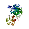
| Title | Structural insights into the processivity of endopolygalacturonase I from Aspergillus niger | ||||||||||||
 Components Components | Polygalacturonase I | ||||||||||||
 Keywords Keywords | HYDROLASE / beta-helix | ||||||||||||
| Function / homology |  Function and homology information Function and homology informationgalacturan 1,4-alpha-galacturonidase activity / endo-polygalacturonase / polygalacturonase activity / pectin catabolic process / cell wall organization / extracellular region Similarity search - Function | ||||||||||||
| Biological species |  | ||||||||||||
| Method |  X-RAY DIFFRACTION / SYNCHROTRON / MOLECULAR REPLACEMENT / Resolution: 1.7 Å X-RAY DIFFRACTION / SYNCHROTRON / MOLECULAR REPLACEMENT / Resolution: 1.7 Å | ||||||||||||
 Authors Authors | van Pouderoyen, G. / Snijder, H.J. / Benen, J.A. / Dijkstra, B.W. | ||||||||||||
 Citation Citation | Journal: Febs Lett. / Year: 2003 Title: Structural insights into the processivity of endopolygalacturonase I from Aspergillus niger. Authors: van Pouderoyen, G. / Snijder, H.J. / Benen, J.A. / Dijkstra, B.W. | ||||||||||||
| History |
|
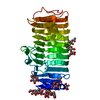
- Structure visualization
| Structure viewer | Molecule: MolmilJmol/JSmol |
|---|
- Downloads & links
-Download
| PDBx/mmCIF format | 1nhc.cif.gz | 407.7 KB | Display | PDBx/mmCIF format |
|---|---|---|---|---|
| PDB format | pdb1nhc.ent.gz | 331.5 KB | Display | PDB format |
| PDBx/mmJSON format | 1nhc.json.gz | Tree view | PDBx/mmJSON format | |
| Others |  Other downloads Other downloads |
-Validation report
| Arichive directory | https://data.pdbj.org/pub/pdb/validation_reports/nh/1nhcftp://data.pdbj.org/pub/pdb/validation_reports/nh/1nhc | HTTPS FTP |
|---|
-Related structure data
| Related structure data |  1czfS S: Starting model for refinement |
|---|---|
| Similar structure data |


-Links
 PDBj
PDBj


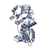
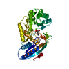
- Assembly
| Deposited unit | 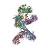
| ||||||||
|---|---|---|---|---|---|---|---|---|---|
| 1 | 
| ||||||||
| 2 | 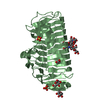
| ||||||||
| 3 | 
| ||||||||
| 4 | 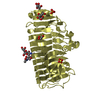
| ||||||||
| 5 | 
| ||||||||
| 6 | 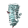
| ||||||||
| Unit cell |
|
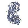
-Components
-Protein , 1 types, 6 molecules ABCDEF
| #1: Protein | Mass: 34891.512 Da / Num. of mol.: 6 Source method: isolated from a genetically manipulated source Source: (gene. exp.) |
|---|
-Sugars , 5 types, 17 molecules 

| #2: Polysaccharide | 2-acetamido-2-deoxy-beta-D-glucopyranose-(1-4)-2-acetamido-2-deoxy-beta-D-glucopyranose Source method: isolated from a genetically manipulated source | ||
|---|---|---|---|
| #3: Polysaccharide | alpha-D-mannopyranose-(1-3)-[alpha-D-mannopyranose-(1-6)]alpha-D-mannopyranose-(1-6)-beta-D- ...alpha-D-mannopyranose-(1-3)-[alpha-D-mannopyranose-(1-6)]alpha-D-mannopyranose-(1-6)-beta-D-mannopyranose-(1-4)-2-acetamido-2-deoxy-beta-D-glucopyranose-(1-4)-2-acetamido-2-deoxy-beta-D-glucopyranose Source method: isolated from a genetically manipulated source | ||
| #4: Polysaccharide | alpha-D-mannopyranose-(1-4)-2-acetamido-2-deoxy-beta-D-glucopyranose-(1-4)-2-acetamido-2-deoxy-beta- ...alpha-D-mannopyranose-(1-4)-2-acetamido-2-deoxy-beta-D-glucopyranose-(1-4)-2-acetamido-2-deoxy-beta-D-glucopyranose Source method: isolated from a genetically manipulated source | ||
| #5: Sugar | ChemComp-MAN /  Type: D-saccharide, alpha linking / Mass: 180.156 Da / Num. of mol.: 11 Type: D-saccharide, alpha linking / Mass: 180.156 Da / Num. of mol.: 11Source method: isolated from a genetically manipulated source Formula: C6H12O6 #8: Sugar |  Type: D-saccharide, beta linking / Mass: 221.208 Da / Num. of mol.: 3 Type: D-saccharide, beta linking / Mass: 221.208 Da / Num. of mol.: 3Source method: isolated from a genetically manipulated source Formula: C8H15NO6 |
-Non-polymers , 3 types, 1374 molecules 


| #6: Chemical | ChemComp-SO4 /  Mass: 96.063 Da / Num. of mol.: 16 / Source method: obtained synthetically / Formula: SO4 Mass: 96.063 Da / Num. of mol.: 16 / Source method: obtained synthetically / Formula: SO4#7: Chemical | ChemComp-GOL /  Mass: 92.094 Da / Num. of mol.: 5 / Source method: obtained synthetically / Formula: C3H8O3 Mass: 92.094 Da / Num. of mol.: 5 / Source method: obtained synthetically / Formula: C3H8O3#9: Water | ChemComp-HOH / | Mass: 18.015 Da / Num. of mol.: 1353 / Source method: isolated from a natural source / Formula: H2O |
|---|
-Details
| Has protein modification | Y |
|---|
-Experimental details
-Experiment
| Experiment | Method: X-RAY DIFFRACTION / Number of used crystals: 1 |
|---|
- Sample preparation
| Crystal | Density Matthews: 2.16 Å3/Da / Density % sol: 43 % | |||||||||||||||||||||||||||||||||||||||||||||||||
|---|---|---|---|---|---|---|---|---|---|---|---|---|---|---|---|---|---|---|---|---|---|---|---|---|---|---|---|---|---|---|---|---|---|---|---|---|---|---|---|---|---|---|---|---|---|---|---|---|---|---|
| Crystal grow | Temperature: 295 K / Method: vapor diffusion, hanging drop / pH: 3 Details: PEG 4000, ammonium sulfate, nikkel sulfate, sodium citrate, pH 3.0, VAPOR DIFFUSION, HANGING DROP, temperature 295K | |||||||||||||||||||||||||||||||||||||||||||||||||
| Crystal grow | *PLUS Method: vapor diffusion, hanging drop | |||||||||||||||||||||||||||||||||||||||||||||||||
| Components of the solutions | *PLUS
|
-Data collection
| Diffraction | Mean temperature: 100 K |
|---|---|
| Diffraction source | Source: SYNCHROTRON / Site: ESRF  / Beamline: ID14-2 / Wavelength: 0.934 Å / Beamline: ID14-2 / Wavelength: 0.934 Å |
| Detector | Type: ADSC QUANTUM 4 / Detector: CCD / Date: Nov 29, 2001 / Details: diamond crystal, Ge crystal and toroidal mirror |
| Radiation | Monochromator: diamond crystal / Protocol: SINGLE WAVELENGTH / Monochromatic (M) / Laue (L): M / Scattering type: x-ray |
| Radiation wavelength | Wavelength: 0.934 Å / Relative weight: 1 |
| Reflection | Resolution: 1.7→37 Å / Num. all: 210531 / Num. obs: 205268 / % possible obs: 97.5 % / Observed criterion σ(F): 0 / Observed criterion σ(I): 0 / Redundancy: 2.1 % / Biso Wilson estimate: 21 Å2 / Rmerge(I) obs: 0.043 / Rsym value: 0.043 / Net I/σ(I): 22.2 |
| Reflection shell | Resolution: 1.7→1.74 Å / Redundancy: 1.7 % / Rmerge(I) obs: 0.384 / Mean I/σ(I) obs: 2.1 / Rsym value: 0.384 / % possible all: 95.6 |
| Reflection | *PLUS Num. measured all: 430145 |
| Reflection shell | *PLUS % possible obs: 95.6 % |
- Processing
| Software |
| |||||||||||||||||||||||||
|---|---|---|---|---|---|---|---|---|---|---|---|---|---|---|---|---|---|---|---|---|---|---|---|---|---|---|
| Refinement | Method to determine structure: MOLECULAR REPLACEMENT Starting model: PDB ENTRY 1CZF Resolution: 1.7→35.81 Å / Isotropic thermal model: ISOTROPIC / Cross valid method: THROUGHOUT / σ(F): 0 / σ(I): 0 / Stereochemistry target values: Engh & Huber
| |||||||||||||||||||||||||
| Displacement parameters | Biso mean: 25 Å2
| |||||||||||||||||||||||||
| Refinement step | Cycle: LAST / Resolution: 1.7→35.81 Å
| |||||||||||||||||||||||||
| Refine LS restraints |
| |||||||||||||||||||||||||
| LS refinement shell | Resolution: 1.7→1.744 Å
| |||||||||||||||||||||||||
| Refinement | *PLUS Lowest resolution: 37 Å | |||||||||||||||||||||||||
| Solvent computation | *PLUS | |||||||||||||||||||||||||
| Displacement parameters | *PLUS | |||||||||||||||||||||||||
| Refine LS restraints | *PLUS
|
