 Movie
Movie Controller


+ Open data

- Basic information
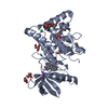
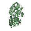
| Entry | Database: PDB / ID: 1ia5 | |||||||||
|---|---|---|---|---|---|---|---|---|---|---|
| Title | POLYGALACTURONASE FROM ASPERGILLUS ACULEATUS | |||||||||
 Components Components | POLYGALACTURONASE | |||||||||
 Keywords Keywords | HYDROLASE / POLYGALACTURONASE / GLYCOSYLHYDROLASE | |||||||||
| Function / homology |  Function and homology information Function and homology informationgalacturan 1,4-alpha-galacturonidase activity / endo-polygalacturonase / polygalacturonase activity / pectin catabolic process / cell wall organization / extracellular region Similarity search - Function | |||||||||
| Biological species |  | |||||||||
| Method |  X-RAY DIFFRACTION / MIR / Resolution: 2 Å X-RAY DIFFRACTION / MIR / Resolution: 2 Å | |||||||||
 Authors Authors | Cho, S.W. / Lee, S. / Shin, W. | |||||||||
 Citation Citation | Journal: J.Mol.Biol. / Year: 2001 Title: The X-ray structure of Aspergillus aculeatus polygalacturonase and a modeled structure of the polygalacturonase-octagalacturonate complex. Authors: Cho, S.W. / Lee, S. / Shin, W. | |||||||||
| History |
|
- Structure visualization
| Structure viewer | Molecule: MolmilJmol/JSmol |
|---|
- Downloads & links
-Download
| PDBx/mmCIF format | 1ia5.cif.gz | 82.1 KB | Display | PDBx/mmCIF format |
|---|---|---|---|---|
| PDB format | pdb1ia5.ent.gz | 61.2 KB | Display | PDB format |
| PDBx/mmJSON format | 1ia5.json.gz | Tree view | PDBx/mmJSON format | |
| Others |  Other downloads Other downloads |
-Validation report
| Arichive directory | https://data.pdbj.org/pub/pdb/validation_reports/ia/1ia5ftp://data.pdbj.org/pub/pdb/validation_reports/ia/1ia5 | HTTPS FTP |
|---|
-Related structure data


-Links
 PDBj
PDBj


- Assembly
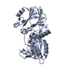
| Deposited unit | 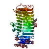
| ||||||||
|---|---|---|---|---|---|---|---|---|---|
| 1 |
| ||||||||
| Unit cell |
|
-Components
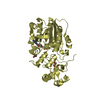
| #1: Protein | Mass: 34675.590 Da / Num. of mol.: 1 / Source method: isolated from a natural source / Source: (natural) References: GenBank: 3220207, UniProt: O74213*PLUS, endo-polygalacturonase | ||||
|---|---|---|---|---|---|
| #2: Polysaccharide | alpha-D-mannopyranose-(1-4)-2-acetamido-2-deoxy-beta-D-glucopyranose-(1-4)-2-acetamido-2-deoxy-beta- ...alpha-D-mannopyranose-(1-4)-2-acetamido-2-deoxy-beta-D-glucopyranose-(1-4)-2-acetamido-2-deoxy-beta-D-glucopyranose Source method: isolated from a genetically manipulated source | ||||
| #3: Sugar | ChemComp-MAN /   Type: D-saccharide, alpha linking / Mass: 180.156 Da / Num. of mol.: 10 Type: D-saccharide, alpha linking / Mass: 180.156 Da / Num. of mol.: 10Source method: isolated from a genetically manipulated source Formula: C6H12O6 #4: Water | ChemComp-HOH / |  Mass: 18.015 Da / Num. of mol.: 193 / Source method: isolated from a natural source / Formula: H2O Mass: 18.015 Da / Num. of mol.: 193 / Source method: isolated from a natural source / Formula: H2OHas protein modification | Y | |
-Experimental details
-Experiment
| Experiment | Method: X-RAY DIFFRACTION / Number of used crystals: 1 |
|---|
- Sample preparation
| Crystal | Density Matthews: 1 Å3/Da / Density % sol: 49.32 % | |||||||||||||||||||||||||
|---|---|---|---|---|---|---|---|---|---|---|---|---|---|---|---|---|---|---|---|---|---|---|---|---|---|---|
| Crystal grow | Temperature: 288 K / Method: vapor diffusion, hanging drop / pH: 8.5 Details: 45% 1,6-HEXANEDIOL, 0.1M TRIS-HCL , 0.2M AMMONIUM ACETATE, pH 8.5, VAPOR DIFFUSION, HANGING DROP, temperature 288K | |||||||||||||||||||||||||
| Crystal grow | *PLUS Method: microdialysis | |||||||||||||||||||||||||
| Components of the solutions | *PLUS
|
-Data collection
| Diffraction | Mean temperature: 288 K |
|---|---|
| Diffraction source | Source: ROTATING ANODE / Type: RIGAKU RU200H / Wavelength: 1.5418 / Wavelength: 1.5418 Å |
| Detector | Type: ENRAF-NONIUS FAST / Detector: AREA DETECTOR / Date: Oct 21, 1999 / Details: COLLIMATOR |
| Radiation | Monochromator: GRAPHITE / Protocol: SINGLE WAVELENGTH / Monochromatic (M) / Laue (L): M / Scattering type: x-ray |
| Radiation wavelength | Wavelength: 1.5418 Å / Relative weight: 1 |
| Reflection | Resolution: 2→50 Å / Num. all: 52967 / Num. obs: 23026 / % possible obs: 85.1 % / Observed criterion σ(F): 2 / Observed criterion σ(I): 4 / Redundancy: 2.3 % / Biso Wilson estimate: 3.3 Å2 / Rmerge(I) obs: 0.056 / Net I/σ(I): 11.5 |
| Reflection shell | Resolution: 2→2.13 Å / Redundancy: 5.3 % / Rmerge(I) obs: 0.124 / % possible all: 81.8 |
| Reflection | *PLUS Highest resolution: 2.02 Å / Lowest resolution: 25 Å / % possible obs: 90.3 % / Num. measured all: 52967 |
| Reflection shell | *PLUS % possible obs: 72.1 % / Rmerge(I) obs: 0.132 |
- Processing
| Software |
| |||||||||||||||||||||||||
|---|---|---|---|---|---|---|---|---|---|---|---|---|---|---|---|---|---|---|---|---|---|---|---|---|---|---|
| Refinement | Method to determine structure: MIR / Resolution: 2→44.74 Å / Cross valid method: THROUGHOUT / σ(F): 0 / σ(I): 0 / Stereochemistry target values: Engh & Huber
| |||||||||||||||||||||||||
| Refine analyze |
| |||||||||||||||||||||||||
| Refinement step | Cycle: LAST / Resolution: 2→44.74 Å
| |||||||||||||||||||||||||
| Refine LS restraints |
| |||||||||||||||||||||||||
| LS refinement shell | Resolution: 2→2.13 Å / Rfactor Rfree error: 0.02
| |||||||||||||||||||||||||
| Software | *PLUS Name: CNS / Classification: refinement | |||||||||||||||||||||||||
| Refinement | *PLUS σ(F): 0 / Rfactor Rfree: 0.213 / Highest resolution: 2.02 Å / Lowest resolution: 25 Å | |||||||||||||||||||||||||
| Solvent computation | *PLUS | |||||||||||||||||||||||||
| Displacement parameters | *PLUS | |||||||||||||||||||||||||
| Refine LS restraints | *PLUS
| |||||||||||||||||||||||||
| LS refinement shell | *PLUS Highest resolution: 2 Å / Rfactor Rfree: 0.267 / Rfactor Rwork: 0.202 |
