 Movie
Movie Controller

[English] 日本語
 Yorodumi
- PDB-7me4: Structure of the extracellular WNT-binding module in Drosophila R... -
+ Open data

- Basic information
| Entry | Database: PDB / ID: 7me4 | ||||||
|---|---|---|---|---|---|---|---|
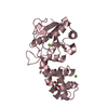
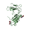
| Title | Structure of the extracellular WNT-binding module in Drosophila Ror2/Nrk | ||||||
 Components Components | Tyrosine-protein kinase transmembrane receptor Ror2 | ||||||
 Keywords Keywords | SIGNALING PROTEIN / WNT signaling / receptor tyrosine kinases / acylation / growth factor signaling / co-receptor / pseudokinases / ligand / receptor / cancer | ||||||
| Function / homology |  Function and homology information Function and homology informationphotoreceptor cell axon guidance / rhabdomere development / Wnt-protein binding / muscle cell cellular homeostasis / transmembrane receptor protein tyrosine kinase activity / cell surface receptor protein tyrosine kinase signaling pathway / receptor protein-tyrosine kinase / positive regulation of neuron projection development / protein tyrosine kinase activity / receptor complex ...photoreceptor cell axon guidance / rhabdomere development / Wnt-protein binding / muscle cell cellular homeostasis / transmembrane receptor protein tyrosine kinase activity / cell surface receptor protein tyrosine kinase signaling pathway / receptor protein-tyrosine kinase / positive regulation of neuron projection development / protein tyrosine kinase activity / receptor complex / positive regulation of phosphatidylinositol 3-kinase/protein kinase B signal transduction / signal transduction / ATP binding / plasma membrane Similarity search - Function | ||||||
| Biological species |  | ||||||
| Method |  X-RAY DIFFRACTION / SYNCHROTRON / MOLECULAR REPLACEMENT / Resolution: 1.75 Å X-RAY DIFFRACTION / SYNCHROTRON / MOLECULAR REPLACEMENT / Resolution: 1.75 Å | ||||||
 Authors Authors | Mendrola, J.M. / Shi, F. / Perry, K. / Stayrook, S.E. / Lemmon, M.A. | ||||||
| Funding support |  United States, 1items United States, 1items
| ||||||
 Citation Citation | Journal: Cell Rep / Year: 2021 Title: ROR and RYK extracellular region structures suggest that receptor tyrosine kinases have distinct WNT-recognition modes. Authors: Shi, F. / Mendrola, J.M. / Sheetz, J.B. / Wu, N. / Sommer, A. / Speer, K.F. / Noordermeer, J.N. / Kan, Z.Y. / Perry, K. / Englander, S.W. / Stayrook, S.E. / Fradkin, L.G. / Lemmon, M.A. | ||||||
| History |
|
- Structure visualization
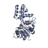
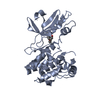
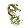
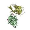
| Structure viewer | Molecule: MolmilJmol/JSmol |
|---|
- Downloads & links
-Download
| PDBx/mmCIF format | 7me4.cif.gz | 125.7 KB | Display | PDBx/mmCIF format |
|---|---|---|---|---|
| PDB format | pdb7me4.ent.gz | 94.2 KB | Display | PDB format |
| PDBx/mmJSON format | 7me4.json.gz | Tree view | PDBx/mmJSON format | |
| Others |  Other downloads Other downloads |
-Validation report
| Summary document | 7me4_validation.pdf.gz | 968.6 KB | Display | wwPDB validaton report |
|---|---|---|---|---|
| Full document | 7me4_full_validation.pdf.gz | 973.1 KB | Display | |
| Data in XML | 7me4_validation.xml.gz | 13.8 KB | Display | |
| Data in CIF | 7me4_validation.cif.gz | 20.3 KB | Display | |
| Arichive directory | https://data.pdbj.org/pub/pdb/validation_reports/me/7me4ftp://data.pdbj.org/pub/pdb/validation_reports/me/7me4 | HTTPS FTP |
-Related structure data
| Related structure data |  7me5C 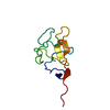 1i71S 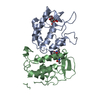 3hklS S: Starting model for refinement C: citing same article ( |
|---|---|
| Similar structure data |


-Links
 PDBj
PDBj


- Assembly
| Deposited unit | 
| ||||||||
|---|---|---|---|---|---|---|---|---|---|
| 1 |
| ||||||||
| Unit cell |
|
-Components
| #1: Protein | Mass: 32587.643 Da / Num. of mol.: 1 / Fragment: Extracellular domain, UNP residues 42-321 Source method: isolated from a genetically manipulated source Source: (gene. exp.)  Spodoptera frugiperda (fall armyworm) Spodoptera frugiperda (fall armyworm)References: UniProt: Q9V6K3, receptor protein-tyrosine kinase |
|---|---|
| #2: Polysaccharide | 2-acetamido-2-deoxy-beta-D-glucopyranose-(1-4)-2-acetamido-2-deoxy-beta-D-glucopyranose Source method: isolated from a genetically manipulated source |
| #3: Chemical | ChemComp-PAM / 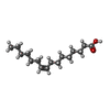  Mass: 254.408 Da / Num. of mol.: 1 / Source method: obtained synthetically / Formula: C16H30O2 Mass: 254.408 Da / Num. of mol.: 1 / Source method: obtained synthetically / Formula: C16H30O2 |
| #4: Water | ChemComp-HOH /  Mass: 18.015 Da / Num. of mol.: 273 / Source method: isolated from a natural source / Formula: H2O Mass: 18.015 Da / Num. of mol.: 273 / Source method: isolated from a natural source / Formula: H2O |
| Has ligand of interest | N |
| Has protein modification | Y |
-Experimental details
-Experiment
| Experiment | Method: X-RAY DIFFRACTION / Number of used crystals: 1 |
|---|
- Sample preparation
| Crystal | Density Matthews: 3.24 Å3/Da / Density % sol: 62.03 % / Description: bar |
|---|---|
| Crystal grow | Temperature: 294.15 K / Method: vapor diffusion, hanging drop / pH: 5 Details: 3 mg/ml protein, 50 mM Bis-Tris propane (pH 5.0), 21% PEG 3350 |
-Data collection
| Diffraction | Mean temperature: 100 K / Serial crystal experiment: N | |||||||||||||||||||||||||||||||||||||||||||||||||||||||||||||||||||||||||||||||||||||||||||||||||||||||||||||||||||||||||
|---|---|---|---|---|---|---|---|---|---|---|---|---|---|---|---|---|---|---|---|---|---|---|---|---|---|---|---|---|---|---|---|---|---|---|---|---|---|---|---|---|---|---|---|---|---|---|---|---|---|---|---|---|---|---|---|---|---|---|---|---|---|---|---|---|---|---|---|---|---|---|---|---|---|---|---|---|---|---|---|---|---|---|---|---|---|---|---|---|---|---|---|---|---|---|---|---|---|---|---|---|---|---|---|---|---|---|---|---|---|---|---|---|---|---|---|---|---|---|---|---|---|---|
| Diffraction source | Source: SYNCHROTRON / Site: APS / Beamline: 24-ID-E / Wavelength: 0.979 Å | |||||||||||||||||||||||||||||||||||||||||||||||||||||||||||||||||||||||||||||||||||||||||||||||||||||||||||||||||||||||||
| Detector | Type: DECTRIS PILATUS 6M-F / Detector: PIXEL / Date: Dec 19, 2012 | |||||||||||||||||||||||||||||||||||||||||||||||||||||||||||||||||||||||||||||||||||||||||||||||||||||||||||||||||||||||||
| Radiation | Protocol: SINGLE WAVELENGTH / Monochromatic (M) / Laue (L): M / Scattering type: x-ray | |||||||||||||||||||||||||||||||||||||||||||||||||||||||||||||||||||||||||||||||||||||||||||||||||||||||||||||||||||||||||
| Radiation wavelength | Wavelength: 0.979 Å / Relative weight: 1 | |||||||||||||||||||||||||||||||||||||||||||||||||||||||||||||||||||||||||||||||||||||||||||||||||||||||||||||||||||||||||
| Reflection | Resolution: 1.75→59.074 Å / Num. all: 36932 / Num. obs: 36932 / % possible obs: 88.5 % / Redundancy: 2.2 % / Rpim(I) all: 0.036 / Rrim(I) all: 0.059 / Rsym value: 0.046 / Net I/av σ(I): 9.4 / Net I/σ(I): 9.1 / Num. measured all: 81880 | |||||||||||||||||||||||||||||||||||||||||||||||||||||||||||||||||||||||||||||||||||||||||||||||||||||||||||||||||||||||||
| Reflection shell | Diffraction-ID: 1
|
- Processing
| Software |
| ||||||||||||||||||||||||||||||||||||||||||||||||||||||||||||||||||||||||||||||||||||||||||||||||||
|---|---|---|---|---|---|---|---|---|---|---|---|---|---|---|---|---|---|---|---|---|---|---|---|---|---|---|---|---|---|---|---|---|---|---|---|---|---|---|---|---|---|---|---|---|---|---|---|---|---|---|---|---|---|---|---|---|---|---|---|---|---|---|---|---|---|---|---|---|---|---|---|---|---|---|---|---|---|---|---|---|---|---|---|---|---|---|---|---|---|---|---|---|---|---|---|---|---|---|---|
| Refinement | Method to determine structure: MOLECULAR REPLACEMENT Starting model: 3HKL 1I71 Resolution: 1.75→42.5 Å / SU ML: 0.23 / Cross valid method: THROUGHOUT / σ(F): 1.34 / Phase error: 26.91 / Stereochemistry target values: ML
| ||||||||||||||||||||||||||||||||||||||||||||||||||||||||||||||||||||||||||||||||||||||||||||||||||
| Solvent computation | Shrinkage radii: 0.9 Å / VDW probe radii: 1.11 Å / Solvent model: FLAT BULK SOLVENT MODEL | ||||||||||||||||||||||||||||||||||||||||||||||||||||||||||||||||||||||||||||||||||||||||||||||||||
| Displacement parameters | Biso max: 116.84 Å2 / Biso mean: 41.7807 Å2 / Biso min: 20.68 Å2 | ||||||||||||||||||||||||||||||||||||||||||||||||||||||||||||||||||||||||||||||||||||||||||||||||||
| Refinement step | Cycle: final / Resolution: 1.75→42.5 Å
| ||||||||||||||||||||||||||||||||||||||||||||||||||||||||||||||||||||||||||||||||||||||||||||||||||
| Refine LS restraints |
| ||||||||||||||||||||||||||||||||||||||||||||||||||||||||||||||||||||||||||||||||||||||||||||||||||
| LS refinement shell | Refine-ID: X-RAY DIFFRACTION / Rfactor Rfree error: 0 / Total num. of bins used: 13
| ||||||||||||||||||||||||||||||||||||||||||||||||||||||||||||||||||||||||||||||||||||||||||||||||||
| Refinement TLS params. | Method: refined / Origin x: 17.4457 Å / Origin y: 33.538 Å / Origin z: -17.8196 Å
| ||||||||||||||||||||||||||||||||||||||||||||||||||||||||||||||||||||||||||||||||||||||||||||||||||
| Refinement TLS group |
|
