 Movie
Movie Controller

[English] 日本語
 Yorodumi
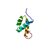
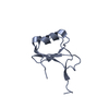
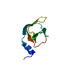
- PDB-1i71: HIGH RESOLUTION CRYSTAL STRUCTURE OF APOLIPOPROTEIN(A) KRINGLE IV... -
+ Open data

- Basic information
| Entry | Database: PDB / ID: 1i71 | ||||||
|---|---|---|---|---|---|---|---|
| Title | HIGH RESOLUTION CRYSTAL STRUCTURE OF APOLIPOPROTEIN(A) KRINGLE IV TYPE 7: INSIGHTS INTO LIGAND BINDING | ||||||
 Components Components | APOLIPOPROTEIN(A) | ||||||
 Keywords Keywords | HYDROLASE / alipoprotein(a) / kringle / protein-ligand interaction / lysine binding | ||||||
| Function / homology |  Function and homology information Function and homology informationplasma lipoprotein particle / LDL remodeling / blood circulation / lipid transport / endopeptidase inhibitor activity / fibronectin binding / Hydrolases; Acting on peptide bonds (peptidases); Serine endopeptidases / apolipoprotein binding / lipid metabolic process / heparin binding ...plasma lipoprotein particle / LDL remodeling / blood circulation / lipid transport / endopeptidase inhibitor activity / fibronectin binding / Hydrolases; Acting on peptide bonds (peptidases); Serine endopeptidases / apolipoprotein binding / lipid metabolic process / heparin binding / serine-type endopeptidase activity / proteolysis / extracellular region Similarity search - Function | ||||||
| Biological species |  Homo sapiens (human) Homo sapiens (human) | ||||||
| Method |  X-RAY DIFFRACTION / SYNCHROTRON / MOLECULAR REPLACEMENT / Resolution: 1.45 Å X-RAY DIFFRACTION / SYNCHROTRON / MOLECULAR REPLACEMENT / Resolution: 1.45 Å | ||||||
 Authors Authors | Ye, Q. / Rahman, M.N. / Koschinsky, M.L. / Jia, Z. | ||||||
 Citation Citation | Journal: Protein Sci. / Year: 2001 Title: High-resolution crystal structure of apolipoprotein(a) kringle IV type 7: insights into ligand binding. Authors: Ye, Q. / Rahman, M.N. / Koschinsky, M.L. / Jia, Z. | ||||||
| History |
|
- Structure visualization
| Structure viewer | Molecule: MolmilJmol/JSmol |
|---|
- Downloads & links
-Download
| PDBx/mmCIF format | 1i71.cif.gz | 31.4 KB | Display | PDBx/mmCIF format |
|---|---|---|---|---|
| PDB format | pdb1i71.ent.gz | 20.5 KB | Display | PDB format |
| PDBx/mmJSON format | 1i71.json.gz | Tree view | PDBx/mmJSON format | |
| Others |  Other downloads Other downloads |
-Validation report
| Arichive directory | https://data.pdbj.org/pub/pdb/validation_reports/i7/1i71ftp://data.pdbj.org/pub/pdb/validation_reports/i7/1i71 | HTTPS FTP |
|---|
-Related structure data
| Related structure data | 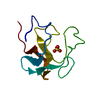 1pk4S S: Starting model for refinement |
|---|---|
| Similar structure data |


-Links
 PDBj
PDBj

- Assembly
| Deposited unit | 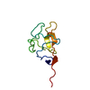
| ||||||||
|---|---|---|---|---|---|---|---|---|---|
| 1 |
| ||||||||
| Unit cell |
|
-Components
| #1: Protein | Mass: 9778.745 Da / Num. of mol.: 1 Fragment: RECOMBINANT KRINGLE IV TYPE 7 (RESIDUES 3781-3863) Source method: isolated from a genetically manipulated source Source: (gene. exp.) Homo sapiens (human) / Plasmid: PET16B / Production host:  References: UniProt: P08519, Hydrolases; Acting on peptide bonds (peptidases); Serine endopeptidases |
|---|---|
| #2: Chemical | ChemComp-SO4 /   Mass: 96.063 Da / Num. of mol.: 1 / Source method: obtained synthetically / Formula: SO4 Mass: 96.063 Da / Num. of mol.: 1 / Source method: obtained synthetically / Formula: SO4 |
| #3: Water | ChemComp-HOH /  Mass: 18.015 Da / Num. of mol.: 116 / Source method: isolated from a natural source / Formula: H2O Mass: 18.015 Da / Num. of mol.: 116 / Source method: isolated from a natural source / Formula: H2O |
| Has protein modification | Y |
-Experimental details
-Experiment
| Experiment | Method: X-RAY DIFFRACTION / Number of used crystals: 1 |
|---|
- Sample preparation
| Crystal | Density Matthews: 2.38 Å3/Da / Density % sol: 48.27 % | |||||||||||||||
|---|---|---|---|---|---|---|---|---|---|---|---|---|---|---|---|---|
| Crystal grow | Temperature: 298 K / Method: vapor diffusion, hanging drop / pH: 5.5 Details: Ammonium sulfate, 2-[N-morpholino]ethane sulfonic acid, , pH 5.5, VAPOR DIFFUSION, HANGING DROP, temperature 298K | |||||||||||||||
| Crystal grow | *PLUS PH range low: 6 / PH range high: 5.5 | |||||||||||||||
| Components of the solutions | *PLUS
|
-Data collection
| Diffraction | Mean temperature: 100 K |
|---|---|
| Diffraction source | Source: SYNCHROTRON / Site: CHESS  / Beamline: A1 / Wavelength: 0.9479 Å / Beamline: A1 / Wavelength: 0.9479 Å |
| Detector | Type: ADSC QUANTUM 4 / Detector: CCD / Date: Jul 20, 2000 / Details: synchrotron optics |
| Radiation | Monochromator: CHESS A1 optics / Protocol: SINGLE WAVELENGTH / Monochromatic (M) / Laue (L): M / Scattering type: x-ray |
| Radiation wavelength | Wavelength: 0.9479 Å / Relative weight: 1 |
| Reflection | Resolution: 1.45→25 Å / Num. all: 48859 / Num. obs: 48859 / % possible obs: 100 % / Observed criterion σ(F): 0 / Observed criterion σ(I): 0 / Redundancy: 2.9 % / Biso Wilson estimate: 15.9 Å2 / Rmerge(I) obs: 0.06 / Rsym value: 0.069 / Net I/σ(I): 14.8 |
| Reflection shell | Resolution: 1.45→1.5 Å / Redundancy: 1.1 % / Rmerge(I) obs: 0.2 / Mean I/σ(I) obs: 2.6 / Num. unique all: 1070 / Rsym value: 0.239 / % possible all: 96.7 |
| Reflection | *PLUS Num. obs: 15774 / % possible obs: 91.8 % / Num. measured all: 48859 / Rmerge(I) obs: 0.069 |
- Processing
| Software |
| |||||||||||||||||||||||||
|---|---|---|---|---|---|---|---|---|---|---|---|---|---|---|---|---|---|---|---|---|---|---|---|---|---|---|
| Refinement | Method to determine structure: MOLECULAR REPLACEMENT Starting model: PDB ENTRY 1PK4 Resolution: 1.45→6 Å / Isotropic thermal model: Isotropic / Cross valid method: THROUGHOUT / σ(F): 0 / σ(I): 0 / Stereochemistry target values: Engh & Huber
| |||||||||||||||||||||||||
| Refinement step | Cycle: LAST / Resolution: 1.45→6 Å
| |||||||||||||||||||||||||
| Refine LS restraints |
| |||||||||||||||||||||||||
| Software | *PLUS Name: CNS / Version: 1 / Classification: refinement | |||||||||||||||||||||||||
| Refinement | *PLUS Lowest resolution: 6 Å / σ(F): 0 / Rfactor obs: 0.171 / Rfactor Rfree: 0.196 | |||||||||||||||||||||||||
| Solvent computation | *PLUS | |||||||||||||||||||||||||
| Displacement parameters | *PLUS | |||||||||||||||||||||||||
| Refine LS restraints | *PLUS
|
