 Movie
Movie Controller

[English] 日本語
 Yorodumi
- PDB-1pk4: CRYSTAL AND MOLECULAR STRUCTURE OF HUMAN PLASMINOGEN KRINGLE 4 RE... -
+ Open data

- Basic information
| Entry | Database: PDB / ID: 1pk4 | ||||||
|---|---|---|---|---|---|---|---|
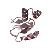
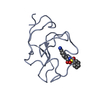
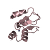
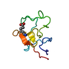
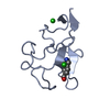
| Title | CRYSTAL AND MOLECULAR STRUCTURE OF HUMAN PLASMINOGEN KRINGLE 4 REFINED AT 1.9-ANGSTROMS RESOLUTION | ||||||
 Components Components | PLASMINOGEN KRINGLE 4 | ||||||
 Keywords Keywords | HYDROLASE(SERINE PROTEASE) | ||||||
| Function / homology |  Function and homology information Function and homology informationplasmin / trans-synaptic signaling by BDNF, modulating synaptic transmission / tissue remodeling / Signaling by PDGF / positive regulation of fibrinolysis / negative regulation of cell-cell adhesion mediated by cadherin / protein antigen binding / Dissolution of Fibrin Clot / biological process involved in interaction with symbiont / Activation of Matrix Metalloproteinases ...plasmin / trans-synaptic signaling by BDNF, modulating synaptic transmission / tissue remodeling / Signaling by PDGF / positive regulation of fibrinolysis / negative regulation of cell-cell adhesion mediated by cadherin / protein antigen binding / Dissolution of Fibrin Clot / biological process involved in interaction with symbiont / Activation of Matrix Metalloproteinases / collagen catabolic process / extracellular matrix disassembly / negative regulation of cell-substrate adhesion / negative regulation of fibrinolysis / apolipoprotein binding / positive regulation of blood vessel endothelial cell migration / fibrinolysis / Degradation of the extracellular matrix / platelet alpha granule lumen / serine-type peptidase activity / Schaffer collateral - CA1 synapse / Regulation of Insulin-like Growth Factor (IGF) transport and uptake by Insulin-like Growth Factor Binding Proteins (IGFBPs) / kinase binding / blood coagulation / Platelet degranulation / protein-folding chaperone binding / extracellular matrix / protease binding / endopeptidase activity / blood microparticle / serine-type endopeptidase activity / external side of plasma membrane / protein domain specific binding / signaling receptor binding / glutamatergic synapse / enzyme binding / cell surface / proteolysis / : / extracellular exosome / extracellular region / plasma membrane Similarity search - Function | ||||||
| Biological species |  Homo sapiens (human) Homo sapiens (human) | ||||||
| Method |  X-RAY DIFFRACTION / Resolution: 1.9 Å X-RAY DIFFRACTION / Resolution: 1.9 Å | ||||||
 Authors Authors | Tulinsky, A. / Mulichak, A.M. | ||||||
 Citation Citation | Journal: Biochemistry / Year: 1991 Title: Crystal and molecular structure of human plasminogen kringle 4 refined at 1.9-A resolution. Authors: Mulichak, A.M. / Tulinsky, A. / Ravichandran, K.G. #1: Journal: J.Mol.Biol. / Year: 1991Title: Structure of Bovine Prothrombin Fragment 1 Refined at 2.25 Angstroms Resolution Authors: Seshadri, T.P. / Tulinsky, A. / Skrzypczak-Jankun, E. / Park, C.H. #2: Journal: Blood Coagulation Fibrinolysis / Year: 1990Title: Structure of the Lysine-Fibrin Binding Subsite of Human Plasminogen Kringle 4 Authors: Mulichak, A.M. / Tulinsky, A. #3: Journal: Proteins / Year: 1988Title: Lysine(Slash)Fibrin Binding Sites of Kringles Modeled After the Structure of Kringle 1 of Prothrombin Authors: Tulinsky, A. / Park, C.H. / Mao, B. / Llinas, M. #4: Journal: J.Mol.Biol. / Year: 1988Title: Structure of Prothrombin Fragment 1 Refined at 2.8 Angstroms Resolution Authors: Tulinsky, A. / Park, C.H. / Skrzypczak-Jankun, E. | ||||||
| History |
|
- Structure visualization
| Structure viewer | Molecule: MolmilJmol/JSmol |
|---|
- Downloads & links
-Download
| PDBx/mmCIF format | 1pk4.cif.gz | 30 KB | Display | PDBx/mmCIF format |
|---|---|---|---|---|
| PDB format | pdb1pk4.ent.gz | 18.7 KB | Display | PDB format |
| PDBx/mmJSON format | 1pk4.json.gz | Tree view | PDBx/mmJSON format | |
| Others |  Other downloads Other downloads |
-Validation report
| Arichive directory | https://data.pdbj.org/pub/pdb/validation_reports/pk/1pk4ftp://data.pdbj.org/pub/pdb/validation_reports/pk/1pk4 | HTTPS FTP |
|---|
-Related structure data
| Similar structure data |
|---|

-Links
 PDBj
PDBj


- Assembly
| Deposited unit | 
| ||||||||
|---|---|---|---|---|---|---|---|---|---|
| 1 |
| ||||||||
| Unit cell |
| ||||||||
| Atom site foot note | 1: CIS PROLINE - PRO 30 |
-Components
| #1: Protein | Mass: 9040.982 Da / Num. of mol.: 1 Source method: isolated from a genetically manipulated source Source: (gene. exp.) Homo sapiens (human) / References: UniProt: P00747 |
|---|---|
| #2: Chemical | ChemComp-SO4 /   Mass: 96.063 Da / Num. of mol.: 1 / Source method: obtained synthetically / Formula: SO4 Mass: 96.063 Da / Num. of mol.: 1 / Source method: obtained synthetically / Formula: SO4 |
| #3: Water | ChemComp-HOH /  Mass: 18.015 Da / Num. of mol.: 96 / Source method: isolated from a natural source / Formula: H2O Mass: 18.015 Da / Num. of mol.: 96 / Source method: isolated from a natural source / Formula: H2O |
| Has protein modification | Y |
-Experimental details
-Experiment
| Experiment | Method: X-RAY DIFFRACTION |
|---|
- Sample preparation
| Crystal | Density Matthews: 2.15 Å3/Da / Density % sol: 42.83 % | ||||||||||||||||||||
|---|---|---|---|---|---|---|---|---|---|---|---|---|---|---|---|---|---|---|---|---|---|
| Crystal grow | *PLUS pH: 6 / Method: vapor diffusion, sitting drop | ||||||||||||||||||||
| Components of the solutions | *PLUS
|
-Data collection
| Radiation | Scattering type: x-ray |
|---|---|
| Radiation wavelength | Relative weight: 1 |
| Reflection | *PLUS Highest resolution: 1.9 Å / Num. all: 6665 / Num. obs: 4869 / % possible obs: 73 % / Observed criterion σ(I): 2 / Rmerge(I) obs: 0.41 |
- Processing
| Software | Name: PROFFT / Classification: refinement | |||||||||||||||||||||||||||||||||||||||||||||||||||||||||||||||
|---|---|---|---|---|---|---|---|---|---|---|---|---|---|---|---|---|---|---|---|---|---|---|---|---|---|---|---|---|---|---|---|---|---|---|---|---|---|---|---|---|---|---|---|---|---|---|---|---|---|---|---|---|---|---|---|---|---|---|---|---|---|---|---|---|
| Refinement | Rfactor obs: 0.142 / Highest resolution: 1.9 Å | |||||||||||||||||||||||||||||||||||||||||||||||||||||||||||||||
| Refinement step | Cycle: LAST / Highest resolution: 1.9 Å
| |||||||||||||||||||||||||||||||||||||||||||||||||||||||||||||||
| Refine LS restraints |
| |||||||||||||||||||||||||||||||||||||||||||||||||||||||||||||||
| Software | *PLUS Name: PROFFT / Classification: refinement | |||||||||||||||||||||||||||||||||||||||||||||||||||||||||||||||
| Refinement | *PLUS Highest resolution: 1.9 Å / Num. reflection obs: 4919 / Rfactor obs: 0.142 | |||||||||||||||||||||||||||||||||||||||||||||||||||||||||||||||
| Solvent computation | *PLUS | |||||||||||||||||||||||||||||||||||||||||||||||||||||||||||||||
| Displacement parameters | *PLUS Biso mean: 18 Å2 | |||||||||||||||||||||||||||||||||||||||||||||||||||||||||||||||
| Refine LS restraints | *PLUS
|
