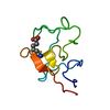
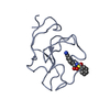
 Movie
Movie Controller


+ Open data

- Basic information
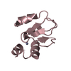
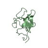
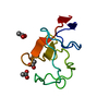
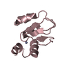
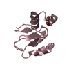
| Entry | Database: PDB / ID: 1kiv | ||||||
|---|---|---|---|---|---|---|---|
| Title | RECOMBINANT KRINGLE IV-10/M66 VARIANT OF HUMAN APOLIPOPROTEIN(A) | ||||||
 Components Components | APOLIPOPROTEIN A | ||||||
 Keywords Keywords | KRINGLE / LYSINE BINDING SITE / APOLIPOPROTEIN(A) | ||||||
| Function / homology |  Function and homology information Function and homology informationplasma lipoprotein particle / LDL remodeling / blood circulation / lipid transport / endopeptidase inhibitor activity / fibronectin binding / Hydrolases; Acting on peptide bonds (peptidases); Serine endopeptidases / apolipoprotein binding / lipid metabolic process / heparin binding ...plasma lipoprotein particle / LDL remodeling / blood circulation / lipid transport / endopeptidase inhibitor activity / fibronectin binding / Hydrolases; Acting on peptide bonds (peptidases); Serine endopeptidases / apolipoprotein binding / lipid metabolic process / heparin binding / serine-type endopeptidase activity / proteolysis / extracellular region Similarity search - Function | ||||||
| Biological species |  Homo sapiens (human) Homo sapiens (human) | ||||||
| Method |  X-RAY DIFFRACTION / MOLECULAR REPLACEMENT / Resolution: 2.1 Å X-RAY DIFFRACTION / MOLECULAR REPLACEMENT / Resolution: 2.1 Å | ||||||
 Authors Authors | Mochalkin, I. / Tulinsky, A. / Scanu, A. | ||||||
 Citation Citation | Journal: Biochemistry / Year: 1999 Title: Recombinant kringle IV-10 modules of human apolipoprotein(a): structure, ligand binding modes, and biological relevance. Authors: Mochalkin, I. / Cheng, B. / Klezovitch, O. / Scanu, A.M. / Tulinsky, A. | ||||||
| History |
|
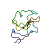
- Structure visualization
| Structure viewer | Molecule: MolmilJmol/JSmol |
|---|
- Downloads & links
-Download
| PDBx/mmCIF format | 1kiv.cif.gz | 30.1 KB | Display | PDBx/mmCIF format |
|---|---|---|---|---|
| PDB format | pdb1kiv.ent.gz | 18.8 KB | Display | PDB format |
| PDBx/mmJSON format | 1kiv.json.gz | Tree view | PDBx/mmJSON format | |
| Others |  Other downloads Other downloads |
-Validation report
| Arichive directory | https://data.pdbj.org/pub/pdb/validation_reports/ki/1kivftp://data.pdbj.org/pub/pdb/validation_reports/ki/1kiv | HTTPS FTP |
|---|
-Related structure data


-Links
 PDBj
PDBj

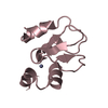
- Assembly
| Deposited unit | 
| ||||||||
|---|---|---|---|---|---|---|---|---|---|
| 1 |
| ||||||||
| Unit cell |
|
-Components
| #1: Protein | Mass: 9070.976 Da / Num. of mol.: 1 / Fragment: KRINGLE IV-10 Source method: isolated from a genetically manipulated source Details: KIV-10/M66 VARIANT OF HUMAN APOLIPOPROTEIN(A) / Source: (gene. exp.) Homo sapiens (human) / Production host:  |
|---|---|
| #2: Water | ChemComp-HOH /  Mass: 18.015 Da / Num. of mol.: 67 / Source method: isolated from a natural source / Formula: H2O Mass: 18.015 Da / Num. of mol.: 67 / Source method: isolated from a natural source / Formula: H2O |
| Compound details | N-TERMINAL (VRQ) AND C-TERMINAL (SDTEGTV) INTERKRINGULAR PARTS ARE DISORDERED. THE P30 RESIDUE IS ...N-TERMINAL (VRQ) AND C-TERMINAL (SDTEGTV) INTERKRING |
| Has protein modification | Y |
-Experimental details
-Experiment
| Experiment | Method: X-RAY DIFFRACTION / Number of used crystals: 1 |
|---|
- Sample preparation
| Crystal | Density Matthews: 1.76 Å3/Da / Density % sol: 30.6 % | ||||||||||||||||||||||||||||||||||||
|---|---|---|---|---|---|---|---|---|---|---|---|---|---|---|---|---|---|---|---|---|---|---|---|---|---|---|---|---|---|---|---|---|---|---|---|---|---|
| Crystal grow | pH: 7 / Details: pH 7.0 | ||||||||||||||||||||||||||||||||||||
| Crystal | *PLUS | ||||||||||||||||||||||||||||||||||||
| Crystal grow | *PLUS Method: vapor diffusion, hanging drop | ||||||||||||||||||||||||||||||||||||
| Components of the solutions | *PLUS
|
-Data collection
| Diffraction | Mean temperature: 130 K |
|---|---|
| Diffraction source | Source: ROTATING ANODE / Type: RIGAKU RUH2R / Wavelength: 1.5418 |
| Detector | Type: RIGAKU RAXIS II / Detector: IMAGE PLATE / Date: Oct 1, 1997 / Details: MSC-YALE MIRRORS |
| Radiation | Monochromator: NI FILTER / Monochromatic (M) / Laue (L): M / Scattering type: x-ray |
| Radiation wavelength | Wavelength: 1.5418 Å / Relative weight: 1 |
| Reflection | Resolution: 2.1→23 Å / Num. obs: 3822 / % possible obs: 81 % / Redundancy: 2.5 % / Rmerge(I) obs: 0.062 / Net I/σ(I): 10.7 |
| Reflection shell | Resolution: 2.1→2.25 Å / Rmerge(I) obs: 0.15 / Mean I/σ(I) obs: 3 / % possible all: 64 |
| Reflection | *PLUS Num. measured all: 9644 |
| Reflection shell | *PLUS % possible obs: 64 % / Rmerge(I) obs: 0.153 |
- Processing
| Software |
| ||||||||||||||||||||||||||||||||||||||||||||||||||||||||||||||||||||||||||||||||||||
|---|---|---|---|---|---|---|---|---|---|---|---|---|---|---|---|---|---|---|---|---|---|---|---|---|---|---|---|---|---|---|---|---|---|---|---|---|---|---|---|---|---|---|---|---|---|---|---|---|---|---|---|---|---|---|---|---|---|---|---|---|---|---|---|---|---|---|---|---|---|---|---|---|---|---|---|---|---|---|---|---|---|---|---|---|---|
| Refinement | Method to determine structure: MOLECULAR REPLACEMENT / Resolution: 2.1→9 Å / σ(F): 1
| ||||||||||||||||||||||||||||||||||||||||||||||||||||||||||||||||||||||||||||||||||||
| Displacement parameters | Biso mean: 21.8 Å2 | ||||||||||||||||||||||||||||||||||||||||||||||||||||||||||||||||||||||||||||||||||||
| Refinement step | Cycle: LAST / Resolution: 2.1→9 Å
| ||||||||||||||||||||||||||||||||||||||||||||||||||||||||||||||||||||||||||||||||||||
| Refine LS restraints |
| ||||||||||||||||||||||||||||||||||||||||||||||||||||||||||||||||||||||||||||||||||||
| Software | *PLUS Name: PROFFT / Classification: refinement | ||||||||||||||||||||||||||||||||||||||||||||||||||||||||||||||||||||||||||||||||||||
| Refinement | *PLUS Rfactor obs: 0.178 | ||||||||||||||||||||||||||||||||||||||||||||||||||||||||||||||||||||||||||||||||||||
| Solvent computation | *PLUS | ||||||||||||||||||||||||||||||||||||||||||||||||||||||||||||||||||||||||||||||||||||
| Displacement parameters | *PLUS |
