 Movie
Movie Controller


+ Open data

- Basic information
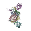
| Entry | Database: PDB / ID: 4j47 | ||||||
|---|---|---|---|---|---|---|---|
| Title | Crystal structure of XIAP-BIR2 domain with SVPI bound | ||||||
 Components Components |
| ||||||
 Keywords Keywords | APOPTOSIS / IAP / XIAP / CASPASE / SMAC / APOPTOSIS INHIBITOR | ||||||
| Function / homology |  Function and homology information Function and homology informationregulation of apoptosis involved in tissue homeostasis / positive regulation of protein linear polyubiquitination / regulation of BMP signaling pathway / copper ion homeostasis / nucleotide-binding oligomerization domain containing 1 signaling pathway / regulation of nucleotide-binding domain, leucine rich repeat containing receptor signaling pathway / nucleotide-binding oligomerization domain containing 2 signaling pathway / SMAC, XIAP-regulated apoptotic response / cysteine-type endopeptidase inhibitor activity involved in apoptotic process / Activation of caspases through apoptosome-mediated cleavage ...regulation of apoptosis involved in tissue homeostasis / positive regulation of protein linear polyubiquitination / regulation of BMP signaling pathway / copper ion homeostasis / nucleotide-binding oligomerization domain containing 1 signaling pathway / regulation of nucleotide-binding domain, leucine rich repeat containing receptor signaling pathway / nucleotide-binding oligomerization domain containing 2 signaling pathway / SMAC, XIAP-regulated apoptotic response / cysteine-type endopeptidase inhibitor activity involved in apoptotic process / Activation of caspases through apoptosome-mediated cleavage / SMAC (DIABLO) binds to IAPs / SMAC(DIABLO)-mediated dissociation of IAP:caspase complexes / Regulation of the apoptosome activity / quinolinate biosynthetic process / TNFR1-induced proapoptotic signaling / RIPK1-mediated regulated necrosis / regulation of innate immune response / cysteine-type endopeptidase inhibitor activity / negative regulation of tumor necrosis factor-mediated signaling pathway / protein K63-linked ubiquitination / positive regulation of type I interferon production / Regulation of PTEN localization / positive regulation of protein ubiquitination / protein serine/threonine kinase binding / TNFR1-induced NF-kappa-B signaling pathway / Deactivation of the beta-catenin transactivating complex / Regulation of TNFR1 signaling / RING-type E3 ubiquitin transferase / positive regulation of JNK cascade / Regulation of necroptotic cell death / Wnt signaling pathway / Regulation of PTEN stability and activity / ubiquitin-protein transferase activity / ubiquitin protein ligase activity / positive regulation of canonical Wnt signaling pathway / regulation of inflammatory response / neuron apoptotic process / regulation of apoptotic process / response to lipopolysaccharide / positive regulation of canonical NF-kappaB signal transduction / defense response to bacterium / regulation of cell cycle / DNA damage response / negative regulation of apoptotic process / zinc ion binding / nucleoplasm / identical protein binding / nucleus / cytoplasm / cytosol Similarity search - Function | ||||||
| Biological species |  Homo sapiens (human)HOMO SAPIENS (human) Homo sapiens (human)HOMO SAPIENS (human) | ||||||
| Method |  X-RAY DIFFRACTION / SYNCHROTRON / FOURIER SYNTHESIS / Resolution: 1.35 Å X-RAY DIFFRACTION / SYNCHROTRON / FOURIER SYNTHESIS / Resolution: 1.35 Å | ||||||
 Authors Authors | Lukacs, C.M. / Janson, C.A. | ||||||
 Citation Citation | Journal: Acta Crystallogr.,Sect.D / Year: 2013 Title: The structure of XIAP BIR2: understanding the selectivity of the BIR domains. Authors: Lukacs, C. / Belunis, C. / Crowther, R. / Danho, W. / Gao, L. / Goggin, B. / Janson, C.A. / Li, S. / Remiszewski, S. / Schutt, A. / Thakur, M.K. / Singh, S.K. / Swaminathan, S. / Pandey, R. ...Authors: Lukacs, C. / Belunis, C. / Crowther, R. / Danho, W. / Gao, L. / Goggin, B. / Janson, C.A. / Li, S. / Remiszewski, S. / Schutt, A. / Thakur, M.K. / Singh, S.K. / Swaminathan, S. / Pandey, R. / Tyagi, R. / Gosu, R. / Kamath, A.V. / Kuglstatter, A. | ||||||
| History |
|
- Structure visualization
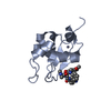
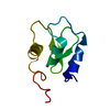
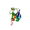
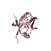
| Structure viewer | Molecule: MolmilJmol/JSmol |
|---|
- Downloads & links
-Download
| PDBx/mmCIF format | 4j47.cif.gz | 83.3 KB | Display | PDBx/mmCIF format |
|---|---|---|---|---|
| PDB format | pdb4j47.ent.gz | 62.3 KB | Display | PDB format |
| PDBx/mmJSON format | 4j47.json.gz | Tree view | PDBx/mmJSON format | |
| Others |  Other downloads Other downloads |
-Validation report
| Arichive directory | https://data.pdbj.org/pub/pdb/validation_reports/j4/4j47ftp://data.pdbj.org/pub/pdb/validation_reports/j4/4j47 | HTTPS FTP |
|---|
-Related structure data
| Related structure data | 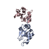 4j3ySC  4j44C  4j45C 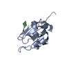 4j46C 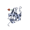 4j48C S: Starting model for refinement C: citing same article ( |
|---|---|
| Similar structure data |


-Links
 PDBj
PDBj


- Assembly
| Deposited unit | 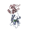
| ||||||||
|---|---|---|---|---|---|---|---|---|---|
| 1 | 
| ||||||||
| 2 | 
| ||||||||
| Unit cell |
|
-Components
| #1: Protein | Mass: 9936.131 Da / Num. of mol.: 2 / Fragment: XIAP-BIR2 RESIDUES 152-236 / Mutation: C202A, C213G Source method: isolated from a genetically manipulated source Source: (gene. exp.) Homo sapiens (human) / Gene: API3, BIRC4, IAP3, U32974, XIAP / Plasmid: PET28A / Production host:  References: UniProt: P98170, Ligases; Forming carbon-nitrogen bonds; Acid-amino-acid ligases (peptide synthases) #2: Protein/peptide | | Mass: 414.496 Da / Num. of mol.: 1 / Source method: obtained synthetically / Details: synthesized / Source: (synth.) HOMO SAPIENS (human)#3: Chemical |   Mass: 65.409 Da / Num. of mol.: 2 / Source method: obtained synthetically / Formula: Zn Mass: 65.409 Da / Num. of mol.: 2 / Source method: obtained synthetically / Formula: Zn#4: Water | ChemComp-HOH / |  Mass: 18.015 Da / Num. of mol.: 149 / Source method: isolated from a natural source / Formula: H2O Mass: 18.015 Da / Num. of mol.: 149 / Source method: isolated from a natural source / Formula: H2O |
|---|
-Experimental details
-Experiment
| Experiment | Method: X-RAY DIFFRACTION / Number of used crystals: 1 |
|---|
- Sample preparation
| Crystal | Density Matthews: 1.86 Å3/Da / Density % sol: 33.88 % |
|---|---|
| Crystal grow | Temperature: 292 K / Method: vapor diffusion, sitting drop / pH: 7 Details: 1.7-1.9 M ammonium sulfate, 125 mM bis-tris propane, pH 7.0, VAPOR DIFFUSION, SITTING DROP, temperature 292K |
-Data collection
| Diffraction | Mean temperature: 100 K |
|---|---|
| Diffraction source | Source: SYNCHROTRON / Site: SLS  / Beamline: X10SA / Beamline: X10SA |
| Detector | Type: DECTRIS PILATUS 6M / Detector: PIXEL / Date: Oct 21, 2012 |
| Radiation | Protocol: SINGLE WAVELENGTH / Monochromatic (M) / Laue (L): M / Scattering type: x-ray |
| Radiation wavelength | Relative weight: 1 |
| Reflection | Resolution: 1.35→38 Å / Num. obs: 33922 / % possible obs: 100 % / Redundancy: 12.5 % / Rmerge(I) obs: 0.051 / Net I/σ(I): 24.3 |
| Reflection shell | Resolution: 1.35→1.42 Å / Redundancy: 12.4 % / Rmerge(I) obs: 0.47 / Mean I/σ(I) obs: 5.5 / % possible all: 100 |
- Processing
| Software |
| ||||||||||||||||||||||||||||||||||||||||||||||||||||||||||||||||||||||||||||||||||||||||||||||||||||||||||||||||||||||||||||||||||||||||||||||||||||||||||||||||||||||||||||||||||||||
|---|---|---|---|---|---|---|---|---|---|---|---|---|---|---|---|---|---|---|---|---|---|---|---|---|---|---|---|---|---|---|---|---|---|---|---|---|---|---|---|---|---|---|---|---|---|---|---|---|---|---|---|---|---|---|---|---|---|---|---|---|---|---|---|---|---|---|---|---|---|---|---|---|---|---|---|---|---|---|---|---|---|---|---|---|---|---|---|---|---|---|---|---|---|---|---|---|---|---|---|---|---|---|---|---|---|---|---|---|---|---|---|---|---|---|---|---|---|---|---|---|---|---|---|---|---|---|---|---|---|---|---|---|---|---|---|---|---|---|---|---|---|---|---|---|---|---|---|---|---|---|---|---|---|---|---|---|---|---|---|---|---|---|---|---|---|---|---|---|---|---|---|---|---|---|---|---|---|---|---|---|---|---|---|
| Refinement | Method to determine structure: FOURIER SYNTHESIS Starting model: 4J3Y Resolution: 1.35→37.27 Å / Cor.coef. Fo:Fc: 0.969 / Cor.coef. Fo:Fc free: 0.959 / SU B: 1.535 / SU ML: 0.038 / Isotropic thermal model: RESTRAINED / Cross valid method: THROUGHOUT / ESU R: 0.057 / ESU R Free: 0.057 / Stereochemistry target values: MAXIMUM LIKELIHOOD / Details: HYDROGENS HAVE BEEN ADDED IN THE RIDING POSITIONS
| ||||||||||||||||||||||||||||||||||||||||||||||||||||||||||||||||||||||||||||||||||||||||||||||||||||||||||||||||||||||||||||||||||||||||||||||||||||||||||||||||||||||||||||||||||||||
| Solvent computation | Ion probe radii: 0.8 Å / Shrinkage radii: 0.8 Å / VDW probe radii: 1.2 Å / Solvent model: MASK | ||||||||||||||||||||||||||||||||||||||||||||||||||||||||||||||||||||||||||||||||||||||||||||||||||||||||||||||||||||||||||||||||||||||||||||||||||||||||||||||||||||||||||||||||||||||
| Displacement parameters | Biso mean: 21.678 Å2
| ||||||||||||||||||||||||||||||||||||||||||||||||||||||||||||||||||||||||||||||||||||||||||||||||||||||||||||||||||||||||||||||||||||||||||||||||||||||||||||||||||||||||||||||||||||||
| Refine analyze |
| ||||||||||||||||||||||||||||||||||||||||||||||||||||||||||||||||||||||||||||||||||||||||||||||||||||||||||||||||||||||||||||||||||||||||||||||||||||||||||||||||||||||||||||||||||||||
| Refinement step | Cycle: LAST / Resolution: 1.35→37.27 Å
| ||||||||||||||||||||||||||||||||||||||||||||||||||||||||||||||||||||||||||||||||||||||||||||||||||||||||||||||||||||||||||||||||||||||||||||||||||||||||||||||||||||||||||||||||||||||
| Refine LS restraints |
|