 Movie
Movie Controller

[English] 日本語
 Yorodumi
- PDB-7lew: Crystal structure of UBE2G2 in complex with the UBE2G2-binding re... -
+ Open data

- Basic information
| Entry | Database: PDB / ID: 7lew | ||||||
|---|---|---|---|---|---|---|---|
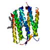
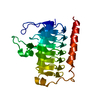
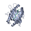
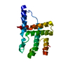
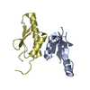
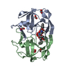
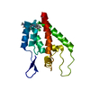
| Title | Crystal structure of UBE2G2 in complex with the UBE2G2-binding region of AUP1 | ||||||
 Components Components |
| ||||||
 Keywords Keywords | LIGASE/Transferase / alpha beta / LIGASE / LIGASE-Transferase complex | ||||||
| Function / homology |  Function and homology information Function and homology informationlipophagy / negative regulation of retrograde protein transport, ER to cytosol / lipid droplet formation / lipid droplet organization / ubiquitin conjugating enzyme binding / protein localization to lipid droplet / retrograde protein transport, ER to cytosol / E2 ubiquitin-conjugating enzyme / ubiquitin conjugating enzyme activity / protein K48-linked ubiquitination ...lipophagy / negative regulation of retrograde protein transport, ER to cytosol / lipid droplet formation / lipid droplet organization / ubiquitin conjugating enzyme binding / protein localization to lipid droplet / retrograde protein transport, ER to cytosol / E2 ubiquitin-conjugating enzyme / ubiquitin conjugating enzyme activity / protein K48-linked ubiquitination / cellular response to interferon-beta / ERAD pathway / lipid droplet / autophagosome / Synthesis of active ubiquitin: roles of E1 and E2 enzymes / ubiquitin binding / response to virus / protein polyubiquitination / ubiquitin-protein transferase activity / Antigen processing: Ubiquitination & Proteasome degradation / cytoplasmic vesicle / ubiquitin-dependent protein catabolic process / ubiquitin protein ligase binding / endoplasmic reticulum membrane / endoplasmic reticulum / extracellular exosome / ATP binding / identical protein binding / membrane / cytosol Similarity search - Function | ||||||
| Biological species |  Homo sapiens (human) Homo sapiens (human) | ||||||
| Method |  X-RAY DIFFRACTION / SYNCHROTRON / MOLECULAR REPLACEMENT / Resolution: 1.736 Å X-RAY DIFFRACTION / SYNCHROTRON / MOLECULAR REPLACEMENT / Resolution: 1.736 Å | ||||||
 Authors Authors | Liang, Y.-H. / Smith, C.E. / Tsai, Y.C. / Weissman, A.M. / Ji, X. | ||||||
 Citation Citation | Journal: Plos Biol. / Year: 2021 Title: A structurally conserved site in AUP1 binds the E2 enzyme UBE2G2 and is essential for ER-associated degradation. Authors: Smith, C.E. / Tsai, Y.C. / Liang, Y.H. / Khago, D. / Mariano, J. / Li, J. / Tarasov, S.G. / Gergel, E. / Tsai, B. / Villaneuva, M. / Clapp, M.E. / Magidson, V. / Chari, R. / Byrd, R.A. / Ji, X. / Weissman, A.M. #1: Journal: Mol Cell / Year: 2009Title: Allosteric activation of E2-RING finger-mediated ubiquitylation by a structurally defined specific E2-binding region of gp78. Authors: Das, R. / Mariano, J. / Tsai, Y.C. / Kalathur, R.C. / Kostova, Z. / Li, J. / Tarasov, S.G. / McFeeters, R.L. / Altieri, A.S. / Ji, X. / Byrd, R.A. / Weissman, A.M. | ||||||
| History |
|
- Structure visualization
| Structure viewer | Molecule: MolmilJmol/JSmol |
|---|
- Downloads & links
-Download
| PDBx/mmCIF format | 7lew.cif.gz | 88.6 KB | Display | PDBx/mmCIF format |
|---|---|---|---|---|
| PDB format | pdb7lew.ent.gz | 65.9 KB | Display | PDB format |
| PDBx/mmJSON format | 7lew.json.gz | Tree view | PDBx/mmJSON format | |
| Others |  Other downloads Other downloads |
-Validation report
| Summary document | 7lew_validation.pdf.gz | 434.8 KB | Display | wwPDB validaton report |
|---|---|---|---|---|
| Full document | 7lew_full_validation.pdf.gz | 436.3 KB | Display | |
| Data in XML | 7lew_validation.xml.gz | 10.8 KB | Display | |
| Data in CIF | 7lew_validation.cif.gz | 15.1 KB | Display | |
| Arichive directory | https://data.pdbj.org/pub/pdb/validation_reports/le/7lewftp://data.pdbj.org/pub/pdb/validation_reports/le/7lew | HTTPS FTP |
-Related structure data
| Related structure data | 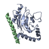 3h8kS S: Starting model for refinement |
|---|---|
| Similar structure data |

-Links
 PDBj
PDBj

- Assembly
| Deposited unit | 
| ||||||||
|---|---|---|---|---|---|---|---|---|---|
| 1 |
| ||||||||
| Unit cell |
|
-Components
| #1: Protein | Mass: 18582.262 Da / Num. of mol.: 1 Source method: isolated from a genetically manipulated source Source: (gene. exp.) Homo sapiens (human) / Gene: UBE2G2, UBC7 / Plasmid: pETDuet-GST / Production host:  References: UniProt: P60604, E2 ubiquitin-conjugating enzyme |
|---|---|
| #2: Protein/peptide | Mass: 4191.585 Da / Num. of mol.: 1 / Fragment: UBE2G2-binding region (G2BR) of AUP1 Source method: isolated from a genetically manipulated source Source: (gene. exp.) Homo sapiens (human) / Gene: AUP1 / Plasmid: pETDuet-GST / Production host: |
| #3: Water | ChemComp-HOH /  Mass: 18.015 Da / Num. of mol.: 158 / Source method: isolated from a natural source / Formula: H2O Mass: 18.015 Da / Num. of mol.: 158 / Source method: isolated from a natural source / Formula: H2O |
-Experimental details
-Experiment
| Experiment | Method: X-RAY DIFFRACTION / Number of used crystals: 1 |
|---|
- Sample preparation
| Crystal | Density Matthews: 2.02 Å3/Da / Density % sol: 38.97 % Description: rod-shaped crystals of dimensions 0.08 mm x 0.08 mm x 0.3 mm |
|---|---|
| Crystal grow | Temperature: 292 K / Method: vapor diffusion, sitting drop / pH: 7.5 / Details: PEG 4000, ammonium acetate etc. |
-Data collection
| Diffraction | Mean temperature: 100 K / Serial crystal experiment: N | |||||||||||||||||||||||||||||||||||||||||||||||||||||||||||||||||||||||||||||
|---|---|---|---|---|---|---|---|---|---|---|---|---|---|---|---|---|---|---|---|---|---|---|---|---|---|---|---|---|---|---|---|---|---|---|---|---|---|---|---|---|---|---|---|---|---|---|---|---|---|---|---|---|---|---|---|---|---|---|---|---|---|---|---|---|---|---|---|---|---|---|---|---|---|---|---|---|---|---|
| Diffraction source | Source: SYNCHROTRON / Site: SSRL  / Beamline: BL9-2 / Wavelength: 0.9794 Å / Beamline: BL9-2 / Wavelength: 0.9794 Å | |||||||||||||||||||||||||||||||||||||||||||||||||||||||||||||||||||||||||||||
| Detector | Type: MARMOSAIC 325 mm CCD / Detector: CCD / Date: Feb 19, 2011 / Details: mirrors | |||||||||||||||||||||||||||||||||||||||||||||||||||||||||||||||||||||||||||||
| Radiation | Monochromator: Double crystal / Protocol: SINGLE WAVELENGTH / Monochromatic (M) / Laue (L): M / Scattering type: x-ray | |||||||||||||||||||||||||||||||||||||||||||||||||||||||||||||||||||||||||||||
| Radiation wavelength | Wavelength: 0.9794 Å / Relative weight: 1 | |||||||||||||||||||||||||||||||||||||||||||||||||||||||||||||||||||||||||||||
| Reflection | Resolution: 1.736→50 Å / Num. obs: 19573 / % possible obs: 99.6 % / Redundancy: 5.4 % / Biso Wilson estimate: 25.15 Å2 / Rmerge(I) obs: 0.034 / Χ2: 1.024 / Net I/σ(I): 16.5 | |||||||||||||||||||||||||||||||||||||||||||||||||||||||||||||||||||||||||||||
| Reflection shell |
|
- Processing
| Software |
| ||||||||||||||||||||||||||||||||||||||||||||||||
|---|---|---|---|---|---|---|---|---|---|---|---|---|---|---|---|---|---|---|---|---|---|---|---|---|---|---|---|---|---|---|---|---|---|---|---|---|---|---|---|---|---|---|---|---|---|---|---|---|---|
| Refinement | Method to determine structure: MOLECULAR REPLACEMENT Starting model: 3H8K Resolution: 1.736→37.819 Å / SU ML: 0.2 / Cross valid method: THROUGHOUT / σ(F): 1.34 / Phase error: 25.12 / Stereochemistry target values: ML
| ||||||||||||||||||||||||||||||||||||||||||||||||
| Solvent computation | Shrinkage radii: 0.9 Å / VDW probe radii: 1.11 Å / Solvent model: FLAT BULK SOLVENT MODEL | ||||||||||||||||||||||||||||||||||||||||||||||||
| Displacement parameters | Biso max: 99.06 Å2 / Biso mean: 36.2691 Å2 / Biso min: 15.05 Å2 | ||||||||||||||||||||||||||||||||||||||||||||||||
| Refinement step | Cycle: final / Resolution: 1.736→37.819 Å
| ||||||||||||||||||||||||||||||||||||||||||||||||
| Refine LS restraints |
| ||||||||||||||||||||||||||||||||||||||||||||||||
| LS refinement shell | Refine-ID: X-RAY DIFFRACTION / Rfactor Rfree error: 0
|
