- PDB-3ery: Different thermodynamic binding mechanisms and peptide fine speci... -
+
Open data
ID or keywords:
Loading...
-
Basic information
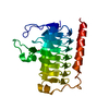
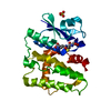
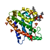
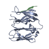
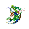
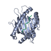
Entry
Database: PDB / ID: 3ery
Title
Different thermodynamic binding mechanisms and peptide fine specificities associated with a panel of structurally similar high-affinity T cell receptors
Components
2-oxoglutarate dehydrogenase E1 peptide
H-2 class I histocompatibility antigen
Keywords
IMMUNE SYSTEM / MHC / Peptide recognition
Function / homology
Function and homology information
olfactory bulb mitral cell layer development / OGDH complex synthesizes succinyl-CoA from 2-OG / 2-oxoglutarate decarboxylation to succinyl-CoA / Glycine degradation / oxoglutarate dehydrogenase (succinyl-transferring) / oxoglutarate dehydrogenase (succinyl-transferring) activity / succinyl-CoA metabolic process / tangential migration from the subventricular zone to the olfactory bulb / cerebellar cortex development / oxoglutarate dehydrogenase complex ...olfactory bulb mitral cell layer development / OGDH complex synthesizes succinyl-CoA from 2-OG / 2-oxoglutarate decarboxylation to succinyl-CoA / Glycine degradation / oxoglutarate dehydrogenase (succinyl-transferring) / oxoglutarate dehydrogenase (succinyl-transferring) activity / succinyl-CoA metabolic process / tangential migration from the subventricular zone to the olfactory bulb / cerebellar cortex development / oxoglutarate dehydrogenase complex / MHC class Ib protein complex / natural killer cell lectin-like receptor binding / TAP2 binding / TAP1 binding / cis-Golgi network membrane / 2-oxoglutarate metabolic process / striatum development / pyramidal neuron development / thalamus development / thiamine pyrophosphate binding / TAP complex binding / Golgi medial cisterna / CD8 receptor binding / beta-2-microglobulin binding / endoplasmic reticulum exit site / TAP binding / MHC class I protein binding / antigen processing and presentation of endogenous peptide antigen via MHC class Ib / antigen processing and presentation of endogenous peptide antigen via MHC class I via ER pathway, TAP-independent / tricarboxylic acid cycle / T cell receptor binding / heat shock protein binding / 14-3-3 protein binding / Mitochondrial protein degradation / glycolytic process / lumenal side of endoplasmic reticulum membrane / hippocampus development / generation of precursor metabolites and energy / defense response / MHC class I peptide loading complex / MHC class I protein complex / positive regulation of T cell mediated cytotoxicity / peptide antigen binding / mitochondrial membrane / phagocytic vesicle membrane / protein-folding chaperone binding / early endosome membrane / early endosome / immune response / mitochondrial matrix / receptor ligand activity / signaling receptor binding / Golgi membrane / external side of plasma membrane / lysosomal membrane / cell surface / Golgi apparatus / endoplasmic reticulum / protein homodimerization activity / mitochondrion / : / metal ion binding / identical protein binding / nucleus / plasma membrane Similarity search - Function
Mass: 1063.202 Da / Num. of mol.: 2 / Source method: obtained synthetically Details: The peptide is chemically synthesized. It is found naturally in human. References: UniProt: Q02218
In the structure databanks used in Yorodumi, some data are registered as the other names, "COVID-19 virus" and "2019-nCoV". Here are the details of the virus and the list of structure data.
Jan 31, 2019. EMDB accession codes are about to change! (news from PDBe EMDB page)
EMDB accession codes are about to change! (news from PDBe EMDB page)
The allocation of 4 digits for EMDB accession codes will soon come to an end. Whilst these codes will remain in use, new EMDB accession codes will include an additional digit and will expand incrementally as the available range of codes is exhausted. The current 4-digit format prefixed with “EMD-” (i.e. EMD-XXXX) will advance to a 5-digit format (i.e. EMD-XXXXX), and so on. It is currently estimated that the 4-digit codes will be depleted around Spring 2019, at which point the 5-digit format will come into force.
The EM Navigator/Yorodumi systems omit the EMD- prefix.
Related info.:Q: What is EMD? / ID/Accession-code notation in Yorodumi/EM Navigator
Yorodumi is a browser for structure data from EMDB, PDB, SASBDB, etc.
This page is also the successor to EM Navigator detail page, and also detail information page/front-end page for Omokage search.
The word "yorodu" (or yorozu) is an old Japanese word meaning "ten thousand". "mi" (miru) is to see.
Related info.:EMDB / PDB / SASBDB / Comparison of 3 databanks / Yorodumi Search / Aug 31, 2016. New EM Navigator & Yorodumi / Yorodumi Papers / Jmol/JSmol / Function and homology information / Changes in new EM Navigator and Yorodumi
 Movie
Movie Controller

 Yorodumi
 Open data
 Basic information
 Components
 Keywords
 Function and homology information
 Homo sapiens (human)
 X-RAY DIFFRACTION /
  Authors
 Citation
 Structure visualization
 Downloads & links
 Other downloads
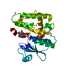


 PDBj
PDBj


 Assembly


 Mass: 18.015 Da / Num. of mol.: 141 / Source method: isolated from a natural source / Formula: H2O
 Sample preparation
 / Beamline: X29A / Wavelength: 0.9785 Å
 Processing