[English] 日本語
Yorodumi
- PDB-4z29: Crystal structure of the magnetobacterial protein MtxA C-terminal... -

+
Open data


ID or keywords:

Loading...

-
Basic information

Entry
Database: PDB / ID: 4z29
TitleCrystal structure of the magnetobacterial protein MtxA C-terminal domain
ComponentsMagnetotaxis protein MtxA
KeywordsSIGNALING PROTEIN / MtxA / TPR / Big
Function / homologyMagnetotaxis protein MtxA
Function and homology information
Biological speciesMagnetospirillum gryphiswaldense (magnetotactic)
MethodX-RAY DIFFRACTION / SYNCHROTRON / SAD / Resolution: 2.03 Å
AuthorsZarivach, R. / Davidov, G.
CitationJournal: Front Mol Biosci / Year: 2015
Title: Crystal structure of the magnetobacterial protein MtxA C-terminal domain reveals a new sequence-structure relationship.
Authors: Davidov, G. / Muller, F.D. / Baumgartner, J. / Bitton, R. / Faivre, D. / Schuler, D. / Zarivach, R.
History
DepositionMar 29, 2015Deposition site: RCSB / Processing site: PDBE
Revision 1.0Jun 3, 2015Provider: repository / Type: Initial release
Revision 1.1Jun 24, 2015Group: Database references

-
Structure visualization

Structure viewerMolecule:
MolmilJmol/JSmol

Downloads & links

-
Assembly

Deposited unit
A: Magnetotaxis protein MtxA
B: Magnetotaxis protein MtxA


Theoretical massNumber of molelcules
Total (without water)69,6442
Polymers69,6442
Non-polymers00
Water2,522140
1
A: Magnetotaxis protein MtxA


Theoretical massNumber of molelcules
Total (without water)34,8221
Polymers34,8221
Non-polymers00
Water181
TypeNameSymmetry operationNumber
identity operation1_555x,y,z1
MethodPISA
2
B: Magnetotaxis protein MtxA


Theoretical massNumber of molelcules
Total (without water)34,8221
Polymers34,8221
Non-polymers00
Water181
TypeNameSymmetry operationNumber
identity operation1_555x,y,z1
MethodPISA
Unit cell
Length a, b, c (Å)40.344, 88.954, 95.400
Angle α, β, γ (deg.)90.000, 90.000, 90.000
Int Tables number19
Space group name H-MP212121
Noncrystallographic symmetry (NCS)NCS domain:
IDEns-IDDetails
11A
21B

NCS domain segments:
Dom-IDComponent-IDEns-IDRefine codeAuth asym-IDAuth seq-ID
1010A137 - 304
2010B137 - 304

-
Components

#1: Protein Magnetotaxis protein MtxA


Mass: 34821.816 Da / Num. of mol.: 2 / Fragment: C-terminal domain, UNP residues 25-313
Source method: isolated from a genetically manipulated source
Source: (gene. exp.) Magnetospirillum gryphiswaldense (magnetotactic)
Gene: mtxA, MGR_0208 / Plasmid: pET-51b(+)Ek/LIC / Production host: Escherichia coli BL21(DE3) (bacteria) / References: UniProt: A4TUL6
#2: Water ChemComp-HOH / water / Water


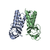
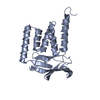
Mass: 18.015 Da / Num. of mol.: 140 / Source method: isolated from a natural source / Formula: H2O

-
Experimental details

-
Experiment

ExperimentMethod: X-RAY DIFFRACTION / Number of used crystals: 1

-
Sample preparation

Crystal growTemperature: 286 K / Method: vapor diffusion, sitting drop / pH: 8.5 / Details: 0.2 M sodium malonate pH 7.0, 25% PEG 3350

-
Data collection

DiffractionMean temperature: 100 K
Diffraction sourceSource: SYNCHROTRON / Site: ESRF / Beamline: ID14-4 / Wavelength: 0.98 Å
DetectorType: ADSC QUANTUM 315r / Detector: CCD / Date: Feb 28, 2013
RadiationProtocol: SINGLE WAVELENGTH / Monochromatic (M) / Laue (L): M / Scattering type: x-ray
Radiation wavelengthWavelength: 0.98 Å / Relative weight: 1
ReflectionResolution: 2.03→50 Å / Num. obs: 22793 / % possible obs: 99.2 % / Redundancy: 11.7 % / Rmerge(I) obs: 0.143 / Χ2: 6.753 / Net I/av σ(I): 44.316 / Net I/σ(I): 12.4 / Num. measured all: 266426
Reflection shell

Diffraction-ID: 1 / Rejects: 0

Resolution (Å)Redundancy (%)Rmerge(I) obsNum. unique allΧ2% possible all
2.03-2.074.90.65910003.64991.3
2.07-2.15.70.60910753.18594.5
2.1-2.146.40.59111102.81498.2
2.14-2.197.20.51311084.95899.7
2.19-2.238.40.44211473.753100
2.23-2.299.80.44311225.711100
2.29-2.3412.40.42311304.906100
2.34-2.4113.80.40211407.767100
2.41-2.48140.34411276.614100
2.48-2.5614.10.32311458.409100
2.56-2.6514.30.29511317.152100
2.65-2.7614.30.25711288.828100
2.76-2.8814.20.22211626.469100
2.88-3.0314.10.18611347.98100
3.03-3.2213.90.14611455.846100
3.22-3.4713.70.12111675.002100
3.47-3.8213.50.10511606.228100
3.82-4.37130.09311816.731100
4.37-5.5112.30.09111968.26499.9
5.51-50120.085128511.2799.8

-
Processing

Software
NameVersionClassification
SCALEPACKdata scaling
REFMAC5.8.0073refinement
PDB_EXTRACT3.15data extraction
HKL-2000data reduction
ARPphasing
RefinementMethod to determine structure: SAD / Resolution: 2.03→47.7 Å / Cor.coef. Fo:Fc: 0.952 / Cor.coef. Fo:Fc free: 0.932 / WRfactor Rfree: 0.2231 / WRfactor Rwork: 0.1819 / FOM work R set: 0.8588 / SU B: 7.993 / SU ML: 0.115 / SU R Cruickshank DPI: 0.2001 / SU Rfree: 0.17 / Cross valid method: THROUGHOUT / σ(F): 0 / ESU R: 0.2 / ESU R Free: 0.17 / Stereochemistry target values: MAXIMUM LIKELIHOOD
Details: HYDROGENS HAVE BEEN ADDED IN THE RIDING POSITIONS U VALUES : WITH TLS ADDED
RfactorNum. reflection% reflectionSelection details
Rfree0.227 1167 5.2 %RANDOM
Rwork0.1848 ---
obs0.1869 21471 98.35 %-
Solvent computationIon probe radii: 0.8 Å / Shrinkage radii: 0.8 Å / VDW probe radii: 1.2 Å / Solvent model: MASK
Displacement parametersBiso max: 137.62 Å2 / Biso mean: 42.506 Å2 / Biso min: 12.43 Å2
Baniso -1Baniso -2Baniso -3
1--0.01 Å20 Å2-0 Å2
2---0.04 Å2-0 Å2
3---0.05 Å2
Refinement stepCycle: final / Resolution: 2.03→47.7 Å
ProteinNucleic acidLigandSolventTotal
Num. atoms2634 0 0 140 2774
Biso mean---33.69 -
Num. residues----345
Refine LS restraints
Refine-IDTypeDev idealDev ideal targetNumber
X-RAY DIFFRACTIONr_bond_refined_d0.0180.0192715
X-RAY DIFFRACTIONr_bond_other_d0.0060.022589
X-RAY DIFFRACTIONr_angle_refined_deg1.8021.9613691
X-RAY DIFFRACTIONr_angle_other_deg1.2435966
X-RAY DIFFRACTIONr_dihedral_angle_1_deg6.1255353
X-RAY DIFFRACTIONr_dihedral_angle_2_deg34.16524.274117
X-RAY DIFFRACTIONr_dihedral_angle_3_deg17.28915455
X-RAY DIFFRACTIONr_dihedral_angle_4_deg22.2531516
X-RAY DIFFRACTIONr_chiral_restr0.0970.2421
X-RAY DIFFRACTIONr_gen_planes_refined0.010.0213098
X-RAY DIFFRACTIONr_gen_planes_other0.0060.02598
X-RAY DIFFRACTIONr_mcbond_it2.1982.4941388
X-RAY DIFFRACTIONr_mcbond_other2.1982.4911387
X-RAY DIFFRACTIONr_mcangle_it3.1953.7181734
Refine LS restraints NCS

Ens-ID: 1 / Number: 9275 / Refine-ID: X-RAY DIFFRACTION / Type: interatomic distance / Rms dev position: 0.13 Å / Weight position: 0.05

Dom-IDAuth asym-ID
1A
2B
LS refinement shellResolution: 2.027→2.079 Å / Total num. of bins used: 20
RfactorNum. reflection% reflection
Rfree0.272 71 -
Rwork0.217 1357 -
all-1428 -
obs--86.44 %
Refinement TLS params.

Method: refined / Refine-ID: X-RAY DIFFRACTION

IDL112)L122)L132)L222)L232)L332)S11 (Å °)S12 (Å °)S13 (Å °)S21 (Å °)S22 (Å °)S23 (Å °)S31 (Å °)S32 (Å °)S33 (Å °)T112)T122)T132)T222)T232)T332)Origin x (Å)Origin y (Å)Origin z (Å)
11.01810.35480.340.72670.11920.90140.0082-0.07510.14290.0747-0.05570.0525-0.0296-0.01490.04750.02480.00230.00430.038-0.02660.031313.987178.515913.2872
21.7186-1.20570.12474.16591.01632.8177-0.01130.03660.1103-0.87910.0404-0.3293-0.62270.2031-0.0290.3301-0.07930.05680.0719-0.03340.074523.375298.467816.2376
Refinement TLS group
IDRefine-IDRefine TLS-IDAuth asym-IDAuth seq-ID
1X-RAY DIFFRACTION1A136 - 311
2X-RAY DIFFRACTION2B137 - 305

+
About Yorodumi

-
News

-
Feb 9, 2022. New format data for meta-information of EMDB entries

New format data for meta-information of EMDB entries

  • Version 3 of the EMDB header file is now the official format.
  • The previous official version 1.9 will be removed from the archive.

Related info.:EMDB header

External links:wwPDB to switch to version 3 of the EMDB data model

-
Aug 12, 2020. Covid-19 info

Covid-19 info

URL: https://pdbj.org/emnavi/covid19.php

New page: Covid-19 featured information page in EM Navigator.

Related info.:Covid-19 info / Mar 5, 2020. Novel coronavirus structure data

+
Mar 5, 2020. Novel coronavirus structure data

Novel coronavirus structure data

Related info.:Yorodumi Speices / Aug 12, 2020. Covid-19 info

External links:COVID-19 featured content - PDBj / Molecule of the Month (242):Coronavirus Proteases

+
Jan 31, 2019. EMDB accession codes are about to change! (news from PDBe EMDB page)

EMDB accession codes are about to change! (news from PDBe EMDB page)

  • The allocation of 4 digits for EMDB accession codes will soon come to an end. Whilst these codes will remain in use, new EMDB accession codes will include an additional digit and will expand incrementally as the available range of codes is exhausted. The current 4-digit format prefixed with “EMD-” (i.e. EMD-XXXX) will advance to a 5-digit format (i.e. EMD-XXXXX), and so on. It is currently estimated that the 4-digit codes will be depleted around Spring 2019, at which point the 5-digit format will come into force.
  • The EM Navigator/Yorodumi systems omit the EMD- prefix.

Related info.:Q: What is EMD? / ID/Accession-code notation in Yorodumi/EM Navigator

External links:EMDB Accession Codes are Changing Soon! / Contact to PDBj

+
Jul 12, 2017. Major update of PDB

Major update of PDB

  • wwPDB released updated PDB data conforming to the new PDBx/mmCIF dictionary.
  • This is a major update changing the version number from 4 to 5, and with Remediation, in which all the entries are updated.
  • In this update, many items about electron microscopy experimental information are reorganized (e.g. em_software).
  • Now, EM Navigator and Yorodumi are based on the updated data.

External links:wwPDB Remediation / Enriched Model Files Conforming to OneDep Data Standards Now Available in the PDB FTP Archive

-
Yorodumi

Thousand views of thousand structures

  • Yorodumi is a browser for structure data from EMDB, PDB, SASBDB, etc.
  • This page is also the successor to EM Navigator detail page, and also detail information page/front-end page for Omokage search.
  • The word "yorodu" (or yorozu) is an old Japanese word meaning "ten thousand". "mi" (miru) is to see.

Related info.:EMDB / PDB / SASBDB / Comparison of 3 databanks / Yorodumi Search / Aug 31, 2016. New EM Navigator & Yorodumi / Yorodumi Papers / Jmol/JSmol / Function and homology information / Changes in new EM Navigator and Yorodumi

Read more