 Movie
Movie Controller

[English] 日本語
 Yorodumi
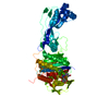
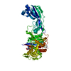
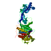
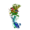
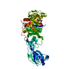
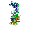
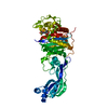
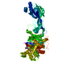
- PDB-7kit: Crystal structure of Pseudomonas aeruginosa PBP3 in complex with ... -
+ Open data

- Basic information
| Entry | Database: PDB / ID: 7kit | ||||||
|---|---|---|---|---|---|---|---|
| Title | Crystal structure of Pseudomonas aeruginosa PBP3 in complex with WCK 4234 | ||||||
 Components Components | Peptidoglycan D,D-transpeptidase FtsI | ||||||
 Keywords Keywords | HYDROLASE/Inhibitor/Antibiotic / Inhibitor / cell wall / antibiotic resistance / HYDROLASE / HYDROLASE-Inhibitor-Antibiotic complex | ||||||
| Function / homology |  Function and homology information Function and homology informationpeptidoglycan glycosyltransferase activity / serine-type D-Ala-D-Ala carboxypeptidase / serine-type D-Ala-D-Ala carboxypeptidase activity / division septum assembly / FtsZ-dependent cytokinesis / penicillin binding / peptidoglycan biosynthetic process / cell wall organization / regulation of cell shape / proteolysis / plasma membrane Similarity search - Function | ||||||
| Biological species |   Pseudomonas aeruginosa (bacteria) Pseudomonas aeruginosa (bacteria) | ||||||
| Method |  X-RAY DIFFRACTION / SYNCHROTRON / MOLECULAR REPLACEMENT / Resolution: 2.089 Å X-RAY DIFFRACTION / SYNCHROTRON / MOLECULAR REPLACEMENT / Resolution: 2.089 Å | ||||||
 Authors Authors | van den Akker, F. | ||||||
 Citation Citation | Journal: Mbio / Year: 2021 Title: Structural Characterization of Diazabicyclooctane beta-Lactam "Enhancers" in Complex with Penicillin-Binding Proteins PBP2 and PBP3 of Pseudomonas aeruginosa. Authors: Rajavel, M. / Kumar, V. / Nguyen, H. / Wyatt, J. / Marshall, S.H. / Papp-Wallace, K.M. / Deshpande, P. / Bhavsar, S. / Yeole, R. / Bhagwat, S. / Patel, M. / Bonomo, R.A. / van den Akker, F. | ||||||
| History |
|
- Structure visualization
| Structure viewer | Molecule: MolmilJmol/JSmol |
|---|
- Downloads & links
-Download
| PDBx/mmCIF format | 7kit.cif.gz | 107.4 KB | Display | PDBx/mmCIF format |
|---|---|---|---|---|
| PDB format | pdb7kit.ent.gz | 79.1 KB | Display | PDB format |
| PDBx/mmJSON format | 7kit.json.gz | Tree view | PDBx/mmJSON format | |
| Others |  Other downloads Other downloads |
-Validation report
| Arichive directory | https://data.pdbj.org/pub/pdb/validation_reports/ki/7kitftp://data.pdbj.org/pub/pdb/validation_reports/ki/7kit | HTTPS FTP |
|---|
-Related structure data
| Related structure data | 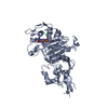 7kisC 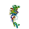 7kivC 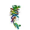 7kiwC 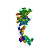 3pbqS S: Starting model for refinement C: citing same article ( |
|---|---|
| Similar structure data |


-Links
 PDBj
PDBj
- Assembly
| Deposited unit | 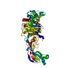
| ||||||||
|---|---|---|---|---|---|---|---|---|---|
| 1 |
| ||||||||
| Unit cell |
|
-Components
| #1: Protein | Mass: 62933.082 Da / Num. of mol.: 1 Source method: isolated from a genetically manipulated source Source: (gene. exp.) Pseudomonas aeruginosa (strain ATCC 15692 / DSM 22644 / CIP 104116 / JCM 14847 / LMG 12228 / 1C / PRS 101 / PAO1) (bacteria)Strain: ATCC 15692 / DSM 22644 / CIP 104116 / JCM 14847 / LMG 12228 / 1C / PRS 101 / PAO1 Gene: ftsI, pbpB, PA4418 / Production host: References: UniProt: G3XD46, serine-type D-Ala-D-Ala carboxypeptidase |
|---|---|
| #2: Chemical | ChemComp-C8Y / (  Mass: 249.244 Da / Num. of mol.: 1 / Source method: obtained synthetically / Formula: C7H11N3O5S / Feature type: SUBJECT OF INVESTIGATION Mass: 249.244 Da / Num. of mol.: 1 / Source method: obtained synthetically / Formula: C7H11N3O5S / Feature type: SUBJECT OF INVESTIGATION |
| #3: Water | ChemComp-HOH /  Mass: 18.015 Da / Num. of mol.: 85 / Source method: isolated from a natural source / Formula: H2O Mass: 18.015 Da / Num. of mol.: 85 / Source method: isolated from a natural source / Formula: H2O |
| Has ligand of interest | Y |
| Has protein modification | Y |
-Experimental details
-Experiment
| Experiment | Method: X-RAY DIFFRACTION / Number of used crystals: 1 |
|---|
- Sample preparation
| Crystal | Density Matthews: 1.83 Å3/Da / Density % sol: 32.76 % |
|---|---|
| Crystal grow | Temperature: 293 K / Method: vapor diffusion, sitting drop Details: 30% polyethylene glycol 4000, 0.2 M MgCl2, 0.1 M Tris pH 8.5 |
-Data collection
| Diffraction | Mean temperature: 100 K / Serial crystal experiment: N |
|---|---|
| Diffraction source | Source: SYNCHROTRON / Site: NSLS  / Beamline: X17B1 / Wavelength: 0.97928 Å / Beamline: X17B1 / Wavelength: 0.97928 Å |
| Detector | Type: DECTRIS EIGER X 16M / Detector: PIXEL / Date: Sep 28, 2019 |
| Radiation | Protocol: SINGLE WAVELENGTH / Monochromatic (M) / Laue (L): M / Scattering type: x-ray |
| Radiation wavelength | Wavelength: 0.97928 Å / Relative weight: 1 |
| Reflection | Resolution: 2.089→29.25 Å / Num. obs: 27862 / % possible obs: 99.4 % / Redundancy: 6.6 % / CC1/2: 0.994 / Rmerge(I) obs: 0.134 / Net I/σ(I): 8.2 |
| Reflection shell | Resolution: 2.089→2.14 Å / Redundancy: 6.2 % / Rmerge(I) obs: 0.92 / Num. unique obs: 1915 / CC1/2: 0.658 / % possible all: 94.3 |
- Processing
| Software |
| ||||||||||||||||||||||||||||||||||||||||||||||||||||||||||||||||||
|---|---|---|---|---|---|---|---|---|---|---|---|---|---|---|---|---|---|---|---|---|---|---|---|---|---|---|---|---|---|---|---|---|---|---|---|---|---|---|---|---|---|---|---|---|---|---|---|---|---|---|---|---|---|---|---|---|---|---|---|---|---|---|---|---|---|---|---|
| Refinement | Method to determine structure: MOLECULAR REPLACEMENT Starting model: 3PBQ Resolution: 2.089→29.249 Å / SU ML: 0.24 / Cross valid method: THROUGHOUT / σ(F): 1.37 / Phase error: 24.14 / Stereochemistry target values: ML
| ||||||||||||||||||||||||||||||||||||||||||||||||||||||||||||||||||
| Solvent computation | Shrinkage radii: 0.9 Å / VDW probe radii: 1.11 Å / Solvent model: FLAT BULK SOLVENT MODEL | ||||||||||||||||||||||||||||||||||||||||||||||||||||||||||||||||||
| Displacement parameters | Biso max: 90.37 Å2 / Biso mean: 37.8607 Å2 / Biso min: 18.18 Å2 | ||||||||||||||||||||||||||||||||||||||||||||||||||||||||||||||||||
| Refinement step | Cycle: final / Resolution: 2.089→29.249 Å
| ||||||||||||||||||||||||||||||||||||||||||||||||||||||||||||||||||
| LS refinement shell | Refine-ID: X-RAY DIFFRACTION / Rfactor Rfree error: 0
|
